What could be more exciting than observing the behavior of microscopic coral in situ? A new underwater microscope from the University of Haifa, part of Israel BioImaging, Euro-BioImaging’s new Node in Israel, allows scientists to study microscopic coral in their native environment – an unprecedented technology development that provides insight into the biology of these fascinating microorganisms - and the small-scale processes that drive large scale ecosystem change.
Taking the microscope out of the lab
Dr. Tali Treibitz, Head of the Marine Imaging Lab, in the Department for Marine Technologies, Charney School of Marine Sciences, University of Haifa, is passionate about the ocean. Trained as a computer scientist and professional diver, she uses her double passion and dual expertise to design complex underwater observation systems to better understand the marine environment. One of the systems developed in her lab is an underwater microscope, a system capable of non-invasively imaging seafloor environments and organisms in situ at nearly micrometre resolution with a computationally extended depth of field.
The microscope was initially developed while Dr. Treibitz was working as a post-doc with Prof. Jules Jaffe in the Jaffe laboratory for Underwater Imaging in the Scripps Institution of Oceanography.
From in vitro to in situ
Designing an instrument capable of imaging microscopic organisms underwater in a dynamic environment presented a number of challenges. These challenges included rapid focusing for dynamic samples, ensuring sufficient illumination for very short exposures, and rapidly adjusting the field of view to image three-dimensional organisms. The development of the imaging system and its applications are described in the Mullen, A. D. et al. article, “Underwater microscopy for in situ studies of benthic ecosystems” published in Nature Communications (2016).
The Benthic Underwater Microscope (BUM) was finalized in 2015. The instrument includes a long working distance objective, an electrically tunable lens (ETL) and focused reflectance illumination. It operates “in situ”, while fully submerged, recording images with an optical resolution of up to 2.2 micrometres. It has a battery capacity of 8h for video and extended time-series recordings and can be left on the reef for autonomous imaging. This enables observation of fine anatomical details of living marine organisms in their natural environment.
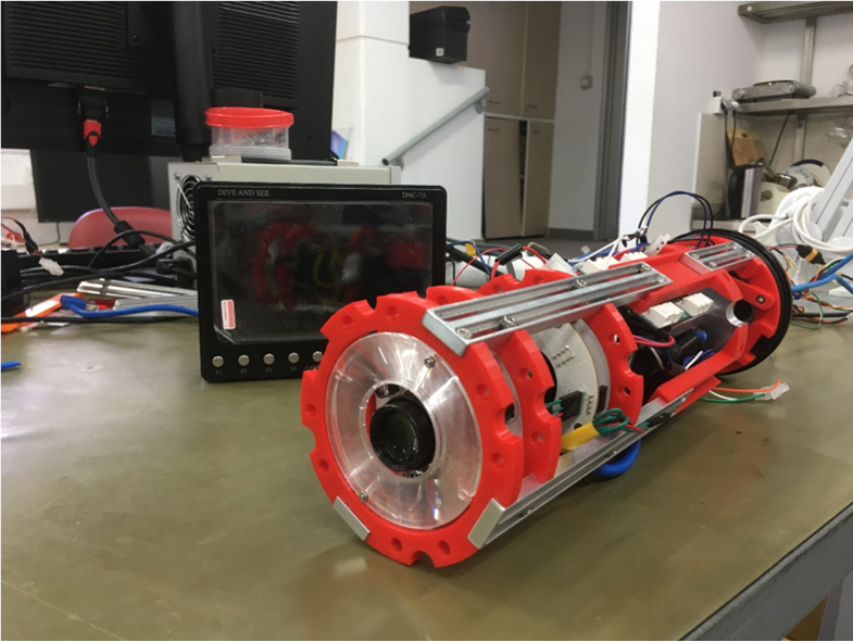
The instrument is made up of one cylindrical aluminum housing and a screen. All of the optical elements are contained in the imaging unit, along with a CCD camera, micro-controller and custom circuit board. The camera’s imaging rate is adjustable – up to 15 frames per second for rapid focal scans to once every several minutes for time-series recordings. The control unit is made up of an on-board computer, 500Gb hard drive for data storage and real-time diver-controlled user interface. The BUM can either be controlled by a diver or via a remotely operated vehicle.
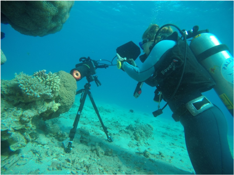
The Benthic Underwater Microscope (BUM) is made up of one cylindrical housing unit and a screen. Photo courtesy of Viseaon Marine Imaging LabThe BUM used to observe coral in its living environment. Photo courtesy of Viseaon Marine Imaging Lab
Small-scale processes – big scale change
Underwater microscopy gives scientists a new way to study benthic marine organisms in their natural environment. This technology has become available at a crucial time, as coral reefs around the world are facing increasing stress due to warmer oceans and increased human activity.
The BUM has been used to examine important biological patterns from a new perspective, the ‘coral polyp’s eye view’. For example, scientists have used the BUM to examine corals experiencing varying levels of bleaching, unveil coral polyp behavior (including feeding and competition), and shed light on algae colonizing the surfaces of living corals. These underwater “in situ” images reveal novel coral behaviors – and are a very important addition to what scientists can see in the lab, or with other systems for macro-level observation – from space, for example.
The most surprising thing that Dr. Treibitz’s team saw in the time lapse movies was that different individual corals from the same species can realize they are from the same species without touching each other, i.e., by chemical signals.
To have a look at some of their beautiful underwater images and get a feel for their discoveries, watch these videos: Coral close-ups and A Seafloor Microscope.
Euro-BioImaging provides open access to biological and biomedical imaging technologies, to all scientists, regardless of their area of expertise or affiliation. Since Dr. Treibitz’s facility at the University of Haifa is part of Israel BioImaging, Euro-BioImaging’s Israeli Node, scientists can apply for open access to this instrument via the Euro-BioImaging website. Knowing how to dive is not a pre-requisite, scientists can also operate a remotely operated vehicle with the microscope attached to it. Training to use the BUM is of course provided. And the microscope can be shipped to study reefs anywhere on the globe.
Read the full article: Mullen, A. D. et al. Underwater microscopy for in situ studies of benthic ecosystems. Nat. Commun. 7:12093 doi: 10.1038/ncomms12093 (2016).
More news from Euro-BioImaging
July 23, 2026
Cellular Imaging Hungary Node expands expertise in advanced neurophotonics
The Euro-BioImaging Cellular Imaging Hungary Node has expanded its service portfolio with the addition of the BrainVisionCenter (BVC) in Budapest. Following a successful…
The UK Euro-BioImaging Node expands from seven to thirteen sites!
We’re delighted to announce that the UK Euro-BioImaging Node is expanding, growing from seven to thirteen sites following a successful upgrade application and…
Building Skills Across Europe: EVOLVE Supports the Second Edition of the Distributed Image Analysis Training Course
The second edition of the “Introduction to Image Analysis with Python for Life Scientists” course marked another successful milestone in the development of Euro-BioImaging’s distributed training model.