
March 30, 2026
Euro-BioImaging at the EOSC ESFRI meeting in Milan
Euro-BioImaging was delighted to attend the ESFRI/EOSC Policy Workshop on “EOSC and Research Infrastructures: Opportunities and Strategies” in Milan from March 15-16, represented by…

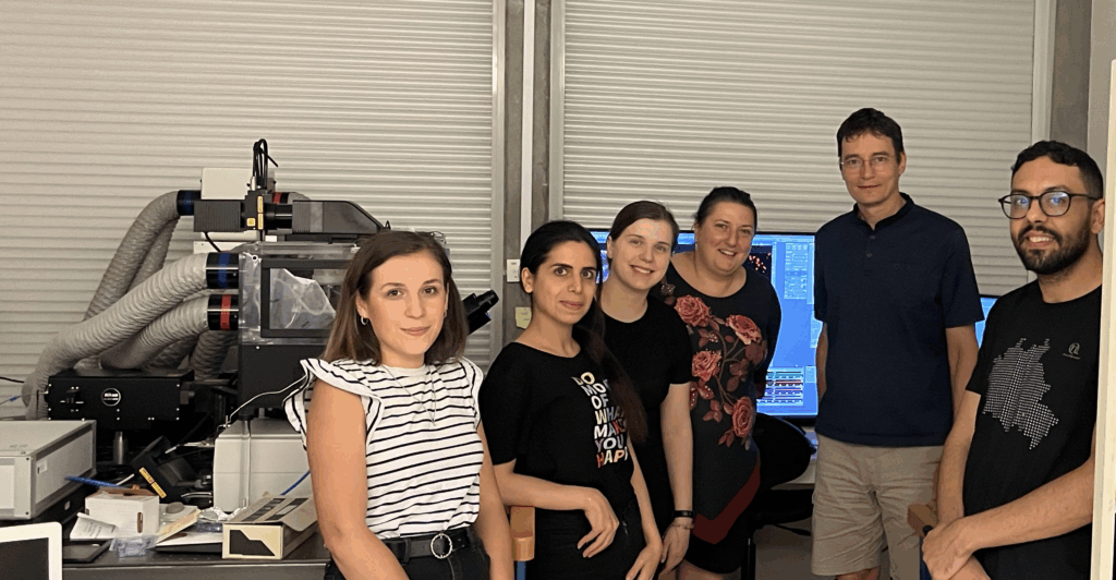
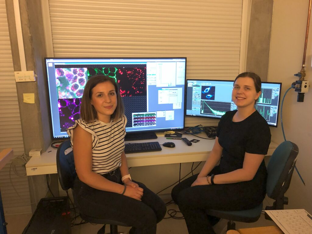
Euro-BioImaging’s Cellular Imaging Hungary Node (University of Debrecen, Hungary) was happy to host Dr. Veronika Huntošová of the Center for Interdisciplinary Biosciences, Technology and Innovation Park at UPJŠ, Slovakia, and her team, for a series of Fluorescence LIfetime Imaging (𝗙𝗟𝗜𝗠) 𝗲𝘅𝗽𝗲𝗿𝗶𝗺𝗲𝗻𝘁𝘀 this summer. The visits were supported by canSERV, which provides cutting-edge cancer research services across Europe and is funded under the Horizon Europe programme under grant agreement number 101058620.
FLIM is an advanced microscopy approach used to explore dynamics in living cells. Supported by the expertise of Dr. Gyorgy Vámosi at the Institute of Biophysics and Cell Biology of the University of Debrecen, part of our Cellular Imaging Hungary Node, Dr. Veronika Huntošová and her team will focus on changes in the endomembrane system of breast cancer cells, using FLIM to monitor them after treatment by DARPin proteins that specifically bind to HER2 receptors of these cells. We are excited to learn more about how this collaboration will contribute to a better understanding of breast cancer!
March 30, 2026
Euro-BioImaging was delighted to attend the ESFRI/EOSC Policy Workshop on “EOSC and Research Infrastructures: Opportunities and Strategies” in Milan from March 15-16, represented by…
March 30, 2026
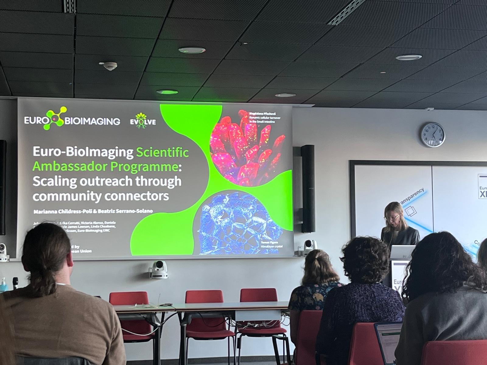
Euro-BioImaging was delighted to attend the Public Awareness & Engagement of Research Infrastructures (PAERI) conference, represented by External Communications Officer, Marianna Childress-Poli. This year’s…

March 26, 2026
The Madrid Advanced Microscopy Center (MAdMiC) is the first Euro-BioImaging Node in Madrid (Spain). It is formed through the collaboration and close work of…