Euro-BioImaging is delighted to share some stories from our fantastic Nodes. First on the long list is the Danish BioImaging Node (DBI). Euro-BioImaging visited the Node and got a two-day tour of their fascinating facilities from Clara Prats, Director of the DBI-INFRA and Director of Light Microscopy and Image Analysis at the University of Copenhagen, and Sonia Garcia, Coordinator of the DBI-INFRA. Clara and Sonia were superb hosts. We filmed what we saw while visiting some of their facilities. Don’t miss our video below and read the article to learn what Danish BioImaging has to offer.
Danish BioImaging is a multi-sited, multimodal Euro-BioImaging Node that brings together five state-of-the-art facilities representing the bioimaging infrastructure of Denmark. The Node provides a broad service offer covering a wide range of advanced bioimaging technologies - from pre-clinical imaging of big animals and humans to cryo-electron microscopy for single particle analysis. Its fields of expertise cover plant biology, HCS of yeast libraries, zoology, pathology, neurosciences and metabolism. In addition, data storage, management and image analysis are a high priority at the DBI Node.
One of the more special features in Danish BioImaging Node is the Bioimaging Core Facility at the Aarhus University Hospital, where surgery rooms are located next to scanners, which Euro-BioImaging users can access and image all kind of animal models and also get access to medical and surgery support. The Danish Molecular Biomedical Imaging Center at the University of Southern Denmark offers some state-of-the-art super resolution methods, such as STED, PALM and single-molecular localization, while also being specialized in home-built systems.
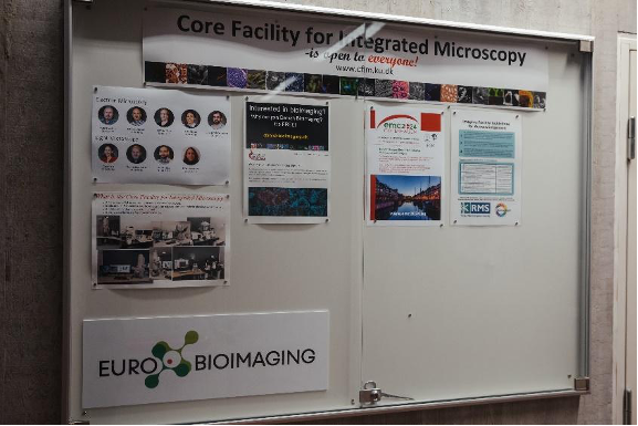
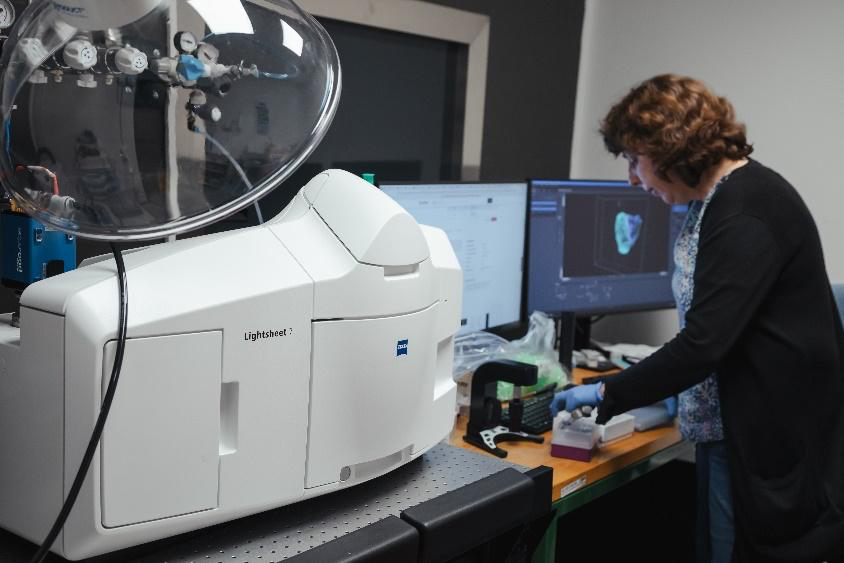
The Center for Advanced BioImaging is specialized in plant biology and yeast. They have a fascinating system up and running; a spinning disk with a vertical stage and with a light controlled software, long-term experiments, such as root growth can be done. The Core Facility for Integrated Microscopy at the University of Copenhagen offers a broad range of applications from electron microscopy to light sheet microscopy with a highly established pathology imaging services with slide scanners, a pathology lab and a combination of deep learning and pixel classification.
On top of all this, the Danish BioImaging Node is very focused on image analysis and are planning to open up image analysis services for people in Denmark and around Europe that will be done in collaboration with computer scientists.
In addition to providing access to advanced imaging technologies, the Danish BioImaging Node is committed to fostering collaboration and knowledge exchange within the scientific community. Through Euro-BioImaging, researchers can request access to the Node's instruments, attend workshops and training courses, and engage in joint research projects. This collaborative atmosphere promotes the rapid dissemination of knowledge and the development of new imaging techniques.
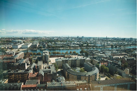
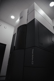
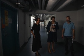
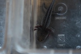
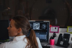
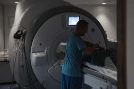
View over Copenhagen from the University of CopenhagenThe Core Facility for Integrated Microscopy with a nice Euro-BioImaging signJana Harizanova inserting samples into the LS7 Light Sheet microscopeThe CM 100 electron microscopeThe facility needed an extra tall room to fit the Titan Krios transmission electron microscopeClara Prats, Sonia Garcia and Michael Pedersen in the maze of the Aarhus University HospitalAn axolotl waiting the get an ultrasound on its heartOngoing patient MRI screeningAnother MRI in use for researchSurgeons and their students during their hands-on course
More news from Euro-BioImaging
July 28, 2026
Global BioImaging Advance the Conversation on Sustainable Biomedical Imaging Facilities
How can biomedical imaging facilities remain scientifically excellent while becoming more financially, environmentally, and operationally sustainable? These questions were at the heart of…
Moara Lemos: New insights from Global BioImaging’s Job Shadowing programme
Moara Lemos is a Brazilian researcher and imaging scientist. Her award winning research pushes the boundaries of structural biology, using advanced cryo-electron microscopy…
A Multiplexed Vision: Highlights from the Special Edition Virtual Pub on Spatial Transcriptomics
On June 12th, Euro-BioImaging hosted its latest Special Edition Virtual Pub, bringing together over 200 researchers and technology developers working at the cutting…