The Euro-BioImaging bookmarks page
Could you guess what was on your Euro-BioImaging bookmark? The answer is here! But that’s not all—dive into our world of cutting-edge imaging, explore our services, and discover the opportunities waiting for you.
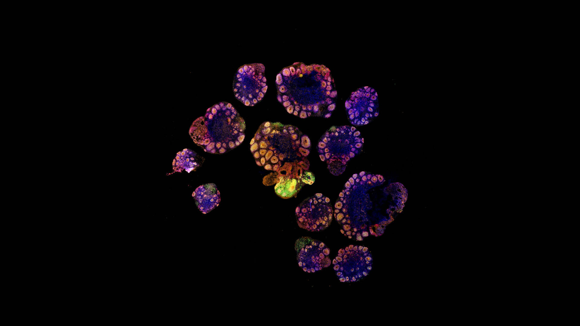
An array of slices of human cortical organoids.
Imaged by Nikky Corthout, VIB Imaging Core Leuven / Sample provided by Franck Maurinot, Pierre Vanderhaeghen lab, VIB Center for Brain and Disease Research

Curly collagen near a breast tumour. This observation provides valuable insight into the tumour microenvironment, potentially influencing tumour behaviour and progression.
Image: Guillaume Jacquemet, Åbo Akademi University
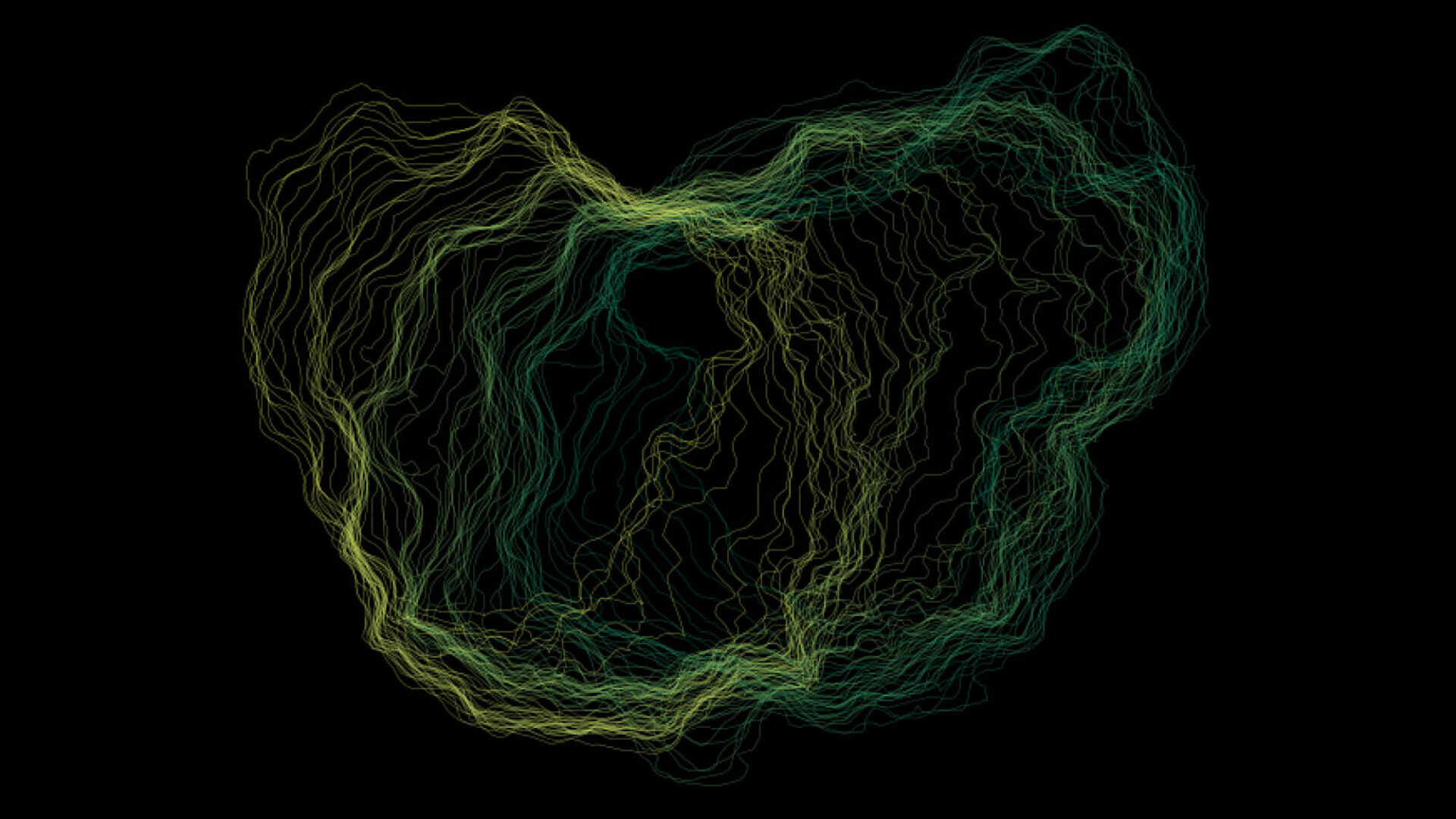
Visualise the evolving outlines of a migrating cancer cell, depicted through a colour gradient representing changes over time. This image captures the dynamic nature of cancer cell movement, revealing insights into their migratory behaviour.
Image: Guillaume Jacquemet, Åbo Akademi University
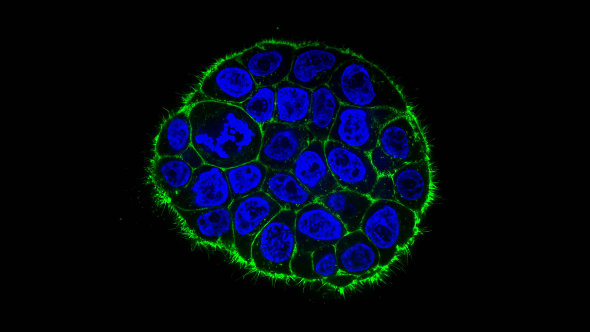
This is a single image capturing the collective migration of breast cancer cells, distinguished by blue nuclei and green actin cytoskeleton. This snapshot unveils the synchronised movement of these cells, providing a glimpse into their invasive potential.
Image: Guillaume Jacquemet, Åbo Akademi University
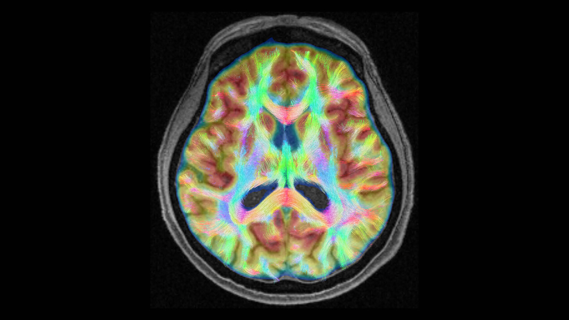
Multimodal view of the human brain. The picture shows an axial slice of the human brain with the fusion of glucose metabolism, structural anatomy and axonal pathways on as revealed, in-vivo, by [18F]-fluorodeoxyglucose positron emission tomography (FDG-PET), T1-weighted magnetic resonance (MR) and diffusion tractography (DT), respectively.
Image: Marco Aiello, NAPLab - IRCCS SDN, Napoli Italy
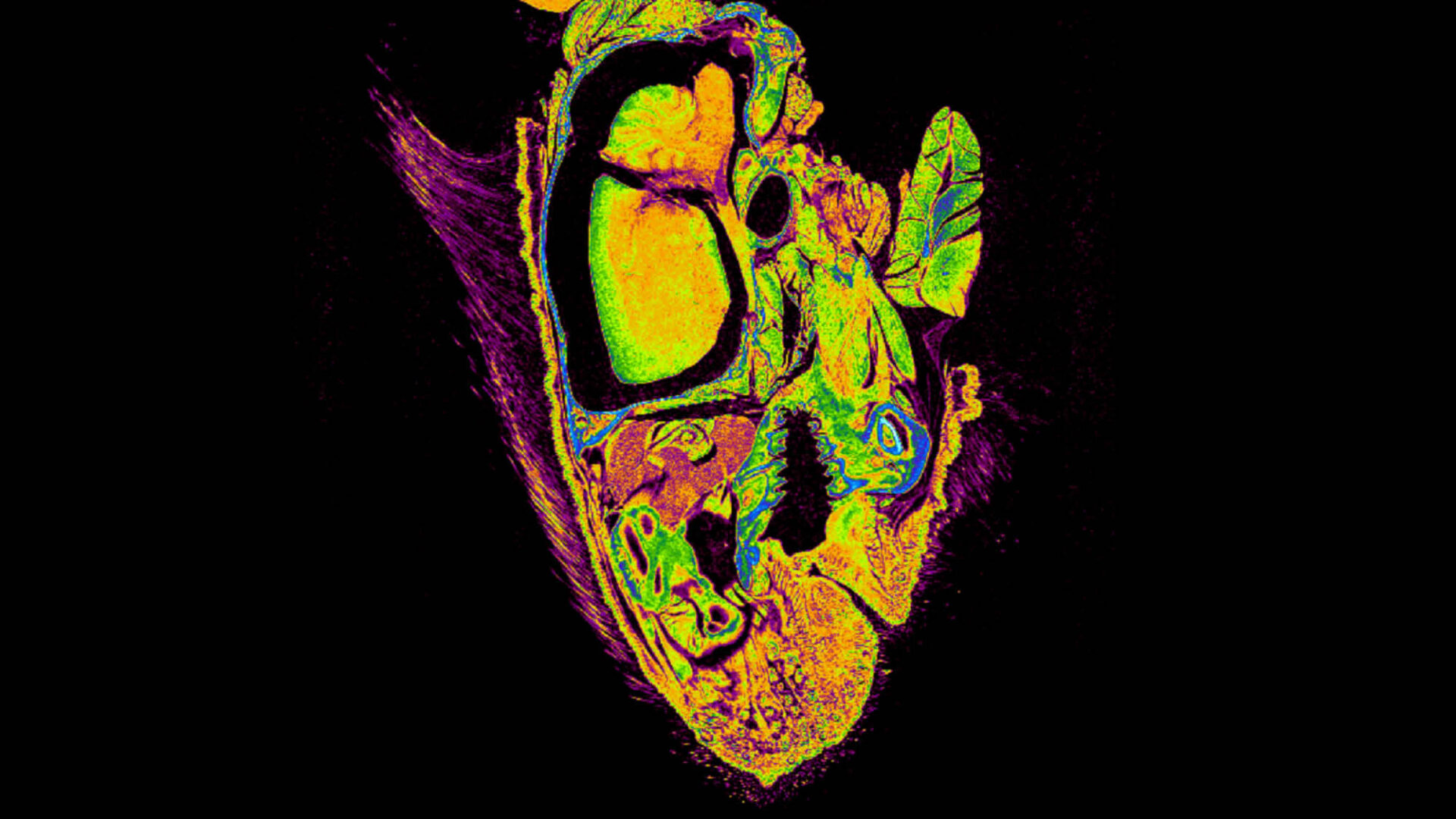
Adult Mouse Head Sagittal Section. A false-colour sagittal section of a dried adult mouse head.
Image: Edgar Alasdair, King’s College, London.
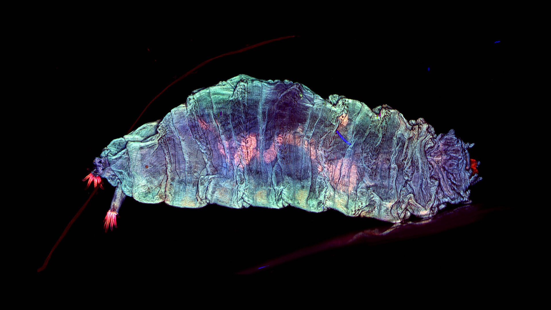
Larva of fruit fly (Drosophila melanogaster) seen through a widefield fluorescent microscope with structured illumination, 5x/0.16 objective and tile scanning method was used with 380 nm, 470 nm, 555 nm and 625 nm LEDs for autofluorescence.
Image: Outi Paloheimo, University of Tampere, Finland.