
March 26, 2026
Welcome, MAdMiC Node!
The Madrid Advanced Microscopy Center (MAdMiC) is the first Euro-BioImaging Node in Madrid (Spain). It is formed through the collaboration and close work of…

Euro-BioImaging is organizing an online User Forum on April 5, 2022, from 14:00-17:00 CEST. This event will highlight the importance of cutting-edge imaging technologies in support of brain research and showcase the specific expertise available at our Nodes across Europe through case studies presented in tandem with the research community.
In this abstract, learn how Joao Castelhano, University of Coimbra, worked alongside the experts at Brain Imaging Network Node in Portugal, to set up a multimodal pipeline with transcranial magnetic stimulation (TMS), magnetic resonance imaging (MRI) and positron emission tomography (PET) to study inter-hemispheric imbalance after unilateral brain damage.
Hear this talk and others like it on April 5 at the Euro-BioImaging User Forum: At the Forefront of Neuroscience.
A TMS and multimodal molecular imaging study of the Interhemispheric inhibition/excitation imbalance: project overview
Joao Castelhano, University of Coimbra
Miguel Castelo-Branco, Brain Imaging Network
The study of the lateralization and imbalances caused by unilateral brain lesion can provide clues on the normal brain lateralization dichotomy. In this context, to further explore this hypothesis of possible inter-hemispheric competition by mutual inhibition a complex set of neuroimaging techniques have to be applied. These include the MRI imaging of structure and function of the brain, as well as PET receptor measures indexing specific inhibitory/excitatory (I/E) processes. Those imaging approaches can be integrated in a multimodal acquisition to study the effects of brain stimulation to understand the Interhemispheric I/E imbalance after unilateral brain damage.
Here, we combine transcranial magnetic stimulation (TMS), magnetic resonance imaging (MRI) and positron emission tomography (PET) to experimentally manipulate the (im)balance between hemispheres which might provide an empirical basis for novel treatment approaches of stroke.
The experiment (19 stroke patients) included two TMS stimulations to experimentally manipulate the (im)balance between hemispheres. Specifically, we target the contralesional hemisphere (left Intra-parietal sulcus (IPS)) with a stimulation protocol that is well-known to decrease cortical excitability (cTBS). Patients receive TMS (placebo and real stimulation) each one followed by one PET 11C-flumazenil measurement to reveal changes of GABA-A receptor binding. The stroke patients also underwent a full Neuropsychological assessment.
All patients performed 3T MRI (anatomical scan, fMRI localizer - a simple saccade paradigm, spectroscopy and resting-state fMRI).
We performed exploratory whole-brain and region-of-interest (ROI)-based analyses (IPS left and right). FMZ data extracted from the whole brain Grey Matter (GM) revealed a significant reduction of GABAa receptors in the right (stroke lesion) hemisphere GM (p<0.0001). Regarding functional MRI data, the TMS stimulation increased brain activity in both hemispheres (p<0.008) and reduced Glx (p=0.009; effect size h2= 0.262).
Our data corroborate the hypothesis of the I/E imbalance that can possibly be recovered by cTBS and might be used as basis to new diagnostic/prognostic biomarkers to be used in neurostimulation studies of brain lesions. This project was performed at Euro-Bioimaging BIN Node in Portugal, within the scope of a Marie Curie project from one external user from the University of Maastricht. The node provides all imaging modalities to perform this complex study.

March 26, 2026
The Madrid Advanced Microscopy Center (MAdMiC) is the first Euro-BioImaging Node in Madrid (Spain). It is formed through the collaboration and close work of…

March 26, 2026
German BioImaging, within its work in the NFDI4BIOIMAGE consortium, and in collaboration with Euro-BioImaging ERIC, has launched a new survey to collect input about…
March 25, 2026
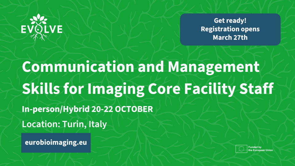
Turin, Italy – 20–22 October 2026 Early career professionals working in imaging core facilities will soon have the opportunity to strengthen essential skills beyond…