A chance to visit our Nodes is always a welcome opportunity for our Hub team members to meet the facility staff, learn more about the institutes that host our Nodes, see their imaging systems, and provide updates on Euro-BioImaging. In June 2024, Johanna Bischof, Head of Bio-Hub operations, got the opportunity to visit the facilities of the Brno Node hosted at CEITEC - MAFIL and CELLIM, where a truly broad range of diverse imaging technologies is available.
From MRI systems for human imaging to phytotrons and lightsheet systems, the Advanced Light Microscopy and Medical Imaging Node Brno is a cutting-edge mixed Node offering open access to its instruments and expertise via Euro-BioImaging.
A visit to the Brno Node
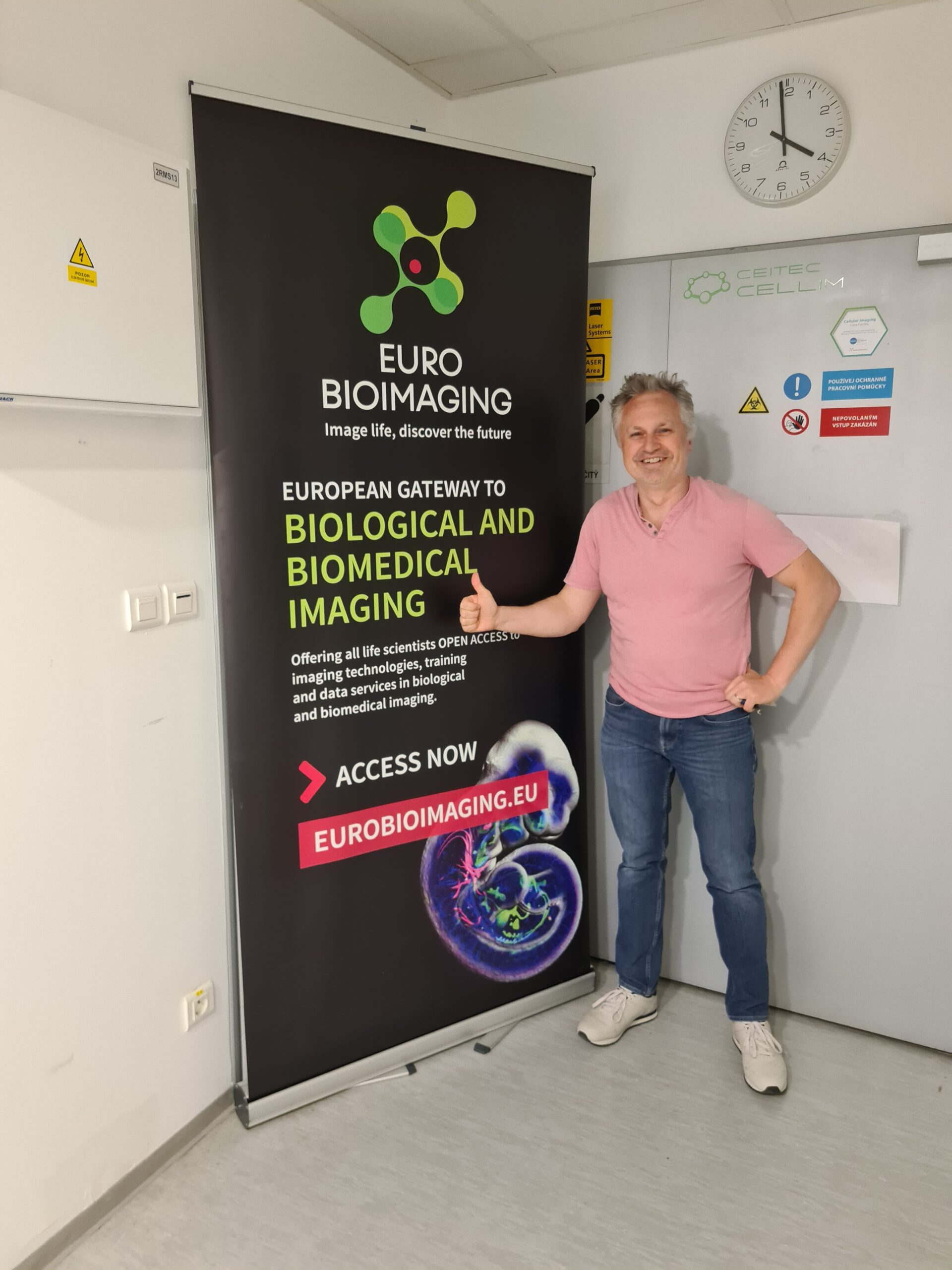
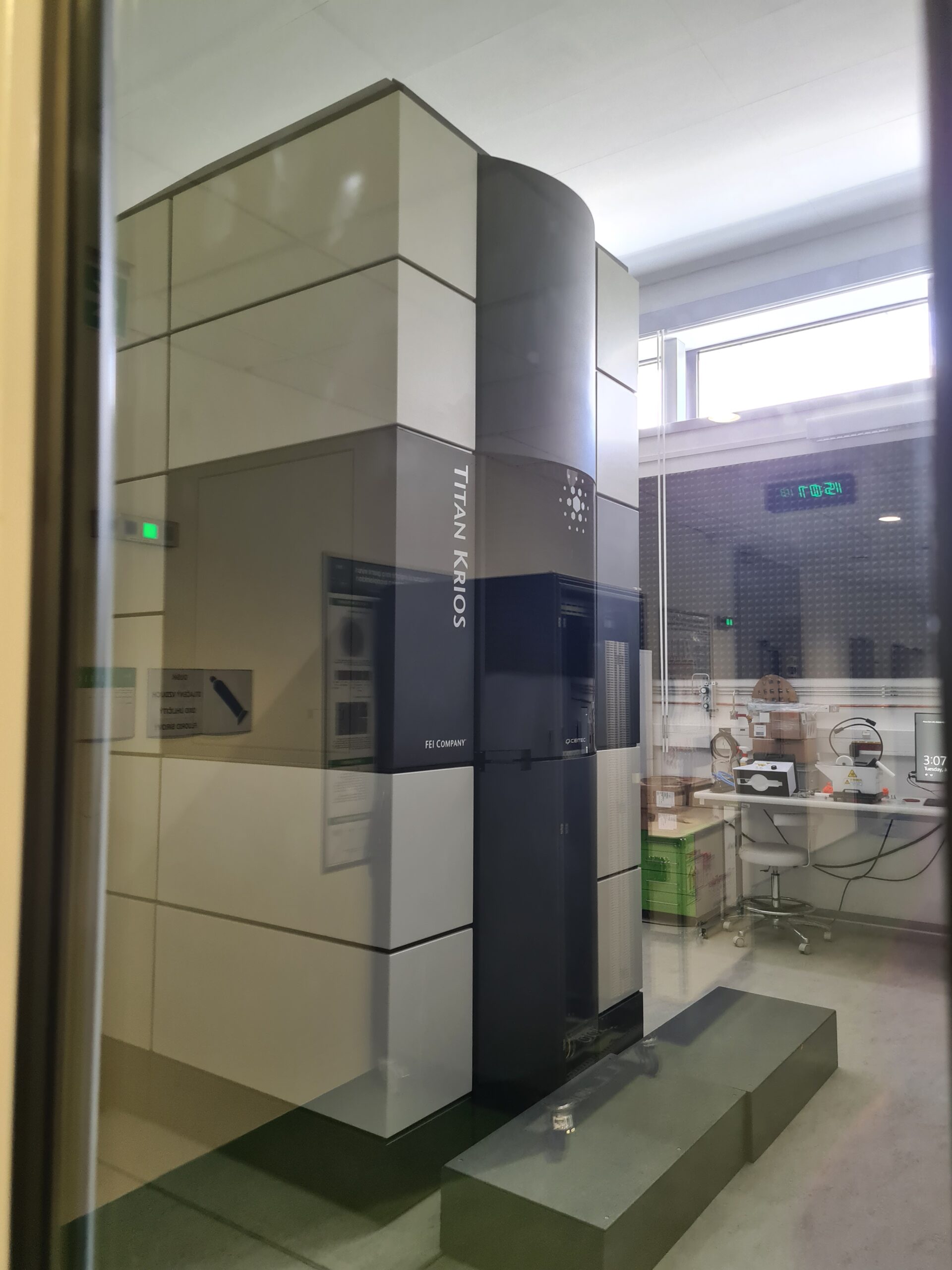
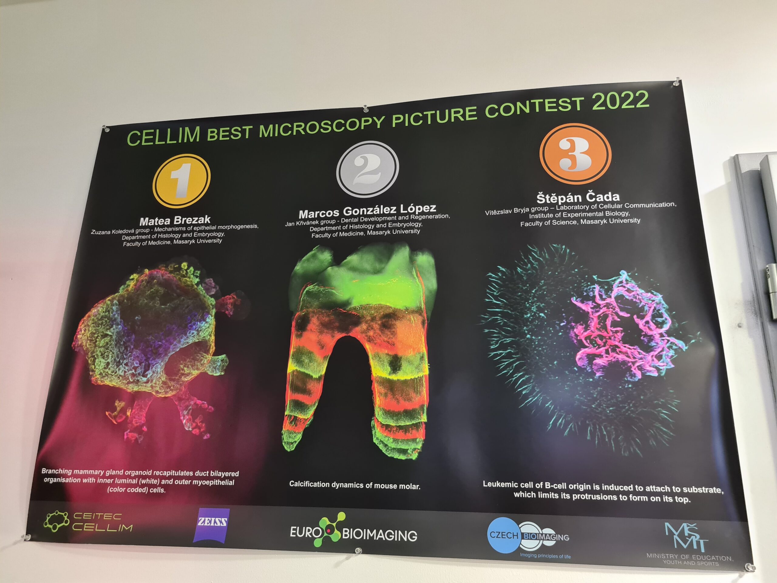
Photo captions from left to right: Milan Esner, Deputy Head of the Brno Node and Representative of microscopic imaging. The Titan KRIOS cryo-EM machine at CEITEC Cellim. Imaging contest winners at CEITEC Cellim. Michal Mikl, Head of the Brno Node and Representative of medical imaging within node. Photos by Johanna Bischof
Medical imaging systems
The MAFIL facility - Multimodal and Functional Imaging Laboratory - headed by Michal MIkl focusses strongly on Neuroscience research and with its two MRI systems allows researchers to study what is going on inside the human brain. EEG - either as a stand-alone or inside the MRI - allows the tracking of brain activity, and the MRI setup of MAFIL also features a range of accessory devices to allow the investigation of different neural processes. These include special auditory and visual input systems as well as keyboards for the volunteers and patients to answer questions while in the MRI - all of course without metal to be compatible with the powerful magnets in the MRI.
Biological imaging systems
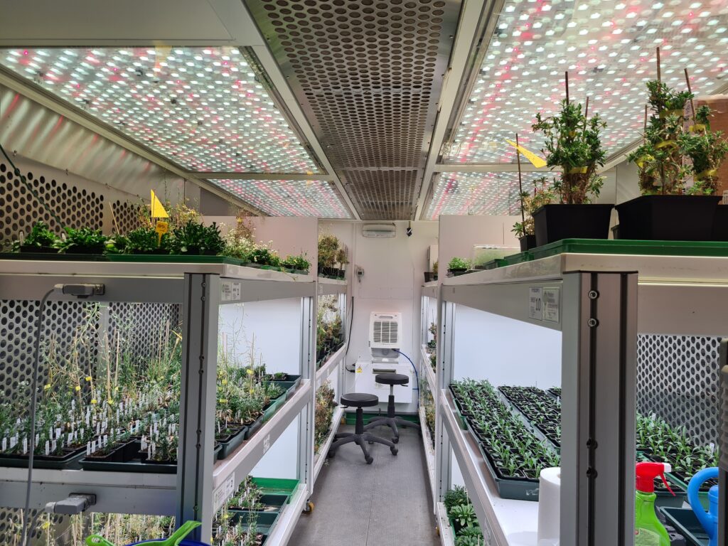
CEITEC - the Central European Institute for Technology - also hosts the CELLIM facility for Cellular Imaging. This facility is headed by Milan Ešner and supports a wide range of different research topics. The first key focus areas are plant biology - where researchers can also take advantage of the institutes' large phytotrons allowing for controlled growth conditions for plants.
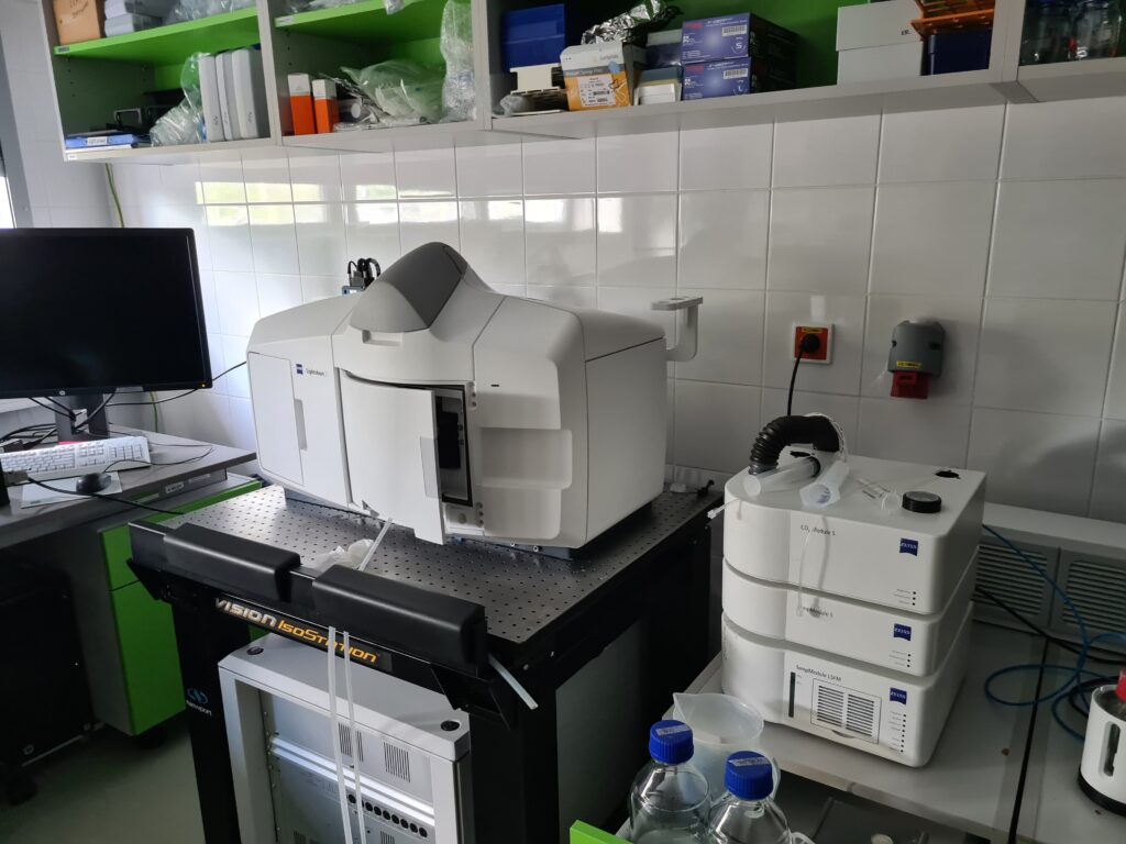
Lightsheet microscope dedicated for imaging of plant samples at the Brno CELLIM facility.
Samples for plant imaging at CEITEC Cellim, part of our Brno Node.
Supporting research on organoid and tissue systems
The second major research area supported by the Node are projects involving organoids and tissue systems. In this area the Node has established a deep expertise on tissue clearing methods and can support researchers not just with the latest lightsheet system to image their organoids but also with needed know-how on how to optimally prepare the samples.
“It is great to see a Node that offers support for research from molecules to humans and the integration and collaboration of the imaging facilities also with the other excellent facilities in the CEITEC institute,”
Johanna Bischof, Head of Bio-Hub Operations, Euro-BioImaging
The expert staff of the facility also supports their users with image analysis and image data management and they are expanding their expertise and service offer in the super-resolution microscopy domain.
Dedicated to service provision
The attention to the user experience and support is a big focus for all the staff in the facility and they are rightfully proud of the amazing pictures they help their users take - displayed throughout the facility and regularly awarded in annual image contests.
“It is great to see a Node that offers support for research from molecules to humans and the integration and collaboration of the imaging facilities also with the other excellent facilities in the CEITEC institute,” says Johanna Bischof.
More news from Euro-BioImaging
July 28, 2026
Global BioImaging Advance the Conversation on Sustainable Biomedical Imaging Facilities
How can biomedical imaging facilities remain scientifically excellent while becoming more financially, environmentally, and operationally sustainable? These questions were at the heart of…
Moara Lemos: New insights from Global BioImaging’s Job Shadowing programme
Moara Lemos is a Brazilian researcher and imaging scientist. Her award winning research pushes the boundaries of structural biology, using advanced cryo-electron microscopy…
A Multiplexed Vision: Highlights from the Special Edition Virtual Pub on Spatial Transcriptomics
On June 12th, Euro-BioImaging hosted its latest Special Edition Virtual Pub, bringing together over 200 researchers and technology developers working at the cutting…