The 8th edition of the From 3D Light to 3D Electron Microscopy symposium, hosted at the Francis Crick Institute in London, brought together a dynamic community of researchers, facility staff, and technology developers working at the forefront of multimodal imaging. The programme reflected the breadth of the field, spanning volume electron microscopy, correlative workflows, X-ray imaging, and data analysis, and highlighted the importance of collaboration across imaging domains. Euro-BioImaging was delighted to attend this community event, represented by Johanna Bischof, Head of Bio-Hub Operations. She shares some highlights from the meeting below.
Strong contributions from Euro-BioImaging Nodes
Euro-BioImaging Nodes were strongly represented throughout the programme, showcasing both technological innovation and practical approaches to making advanced imaging accessible.
In the opening session on Correlative Microscopy – back to basics, several Node experts highlighted how robust workflows can be developed using accessible tools. Ana Laura Vinagre Costa e Sousa (PPBI Node) presented the Histo-CLEM approach, demonstrating how widely available histological techniques can be integrated into correlative workflows to bridge macro- and nanoscale imaging. Her work emphasised that high-quality correlative imaging does not always require highly specialised equipment, but can be achieved through thoughtful workflow design and optimisation.
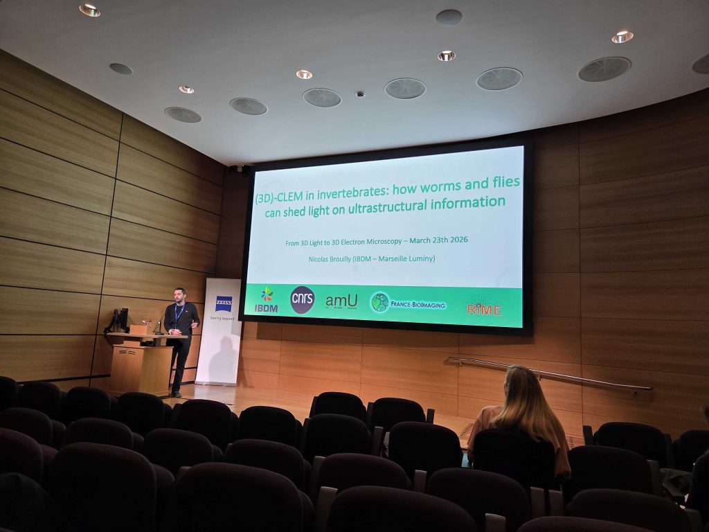
Nicolas Brouilly (France-BioImaging Node) explored complementary CLEM strategies in biological model systems, combining fluorescence imaging, electron microscopy, and tomography to investigate ultrastructural features in organisms such as C. elegans and Drosophila. By comparing pre- and post-embedding approaches, his work highlighted how different methodologies can provide distinct but complementary biological insights, reinforcing the importance of flexible, application-driven workflows.
Nicolas Brouilly, France-BioImaging, presents advances in CLEM at the 3D CLEM meeting in 2026.
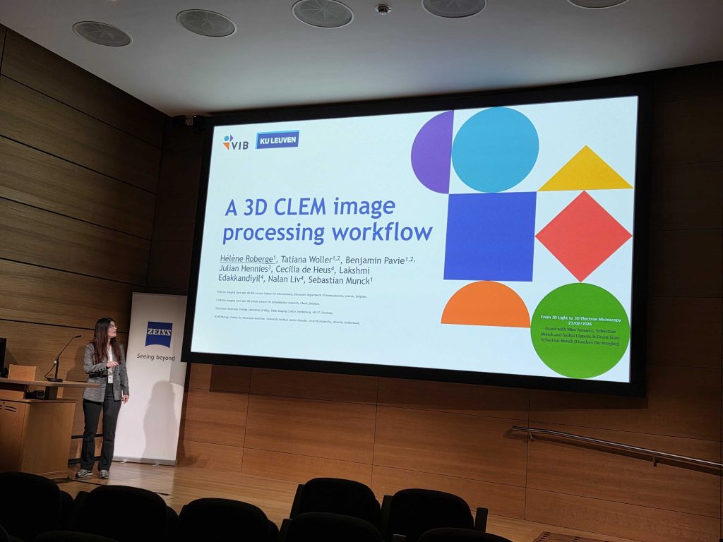
From the Flanders BioImaging Node, Hélène Roberge presented a scalable, open-source image processing pipeline for 3D CLEM datasets. Addressing a key bottleneck in the field, her work integrates tools for alignment, registration, segmentation, and visualisation into a coherent and reproducible workflow. By making these tools modular and openly available, the approach supports both accessibility and reproducibility in handling complex multimodal datasets.
Hélène Roberge (FlaBI Node) presents an open source pipeline for 3D CLEM image analysis.
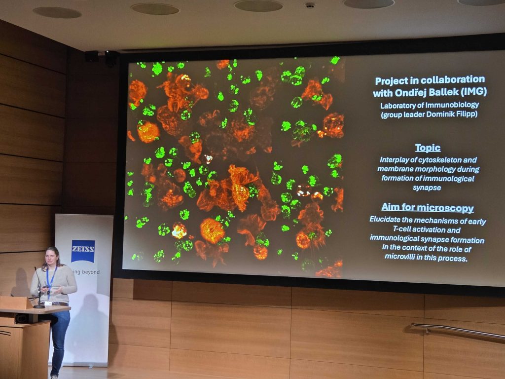
Marketa Dalecká (Prague Node) presented an advanced correlative workflow combining cryo-fluorescence microscopy with FIB-SEM tomography to visualise T-cell microvilli and their role in the immunological synapse. By integrating precise targeting of regions of interest under cryogenic conditions with high-resolution 3D electron microscopy, this approach enables detailed reconstruction of complex cell–cell interfaces. The work highlights how cutting-edge correlative techniques can provide new insights into immune cell interactions while also demonstrating robust, facility-ready workflows for nanoscale imaging
Marketa Dalecká from our Prague Node present FIB-SEM methods at the 3D CLEM meeting 2026.
These contributions reflect how Euro-BioImaging Nodes not only push technological boundaries, but also to ensure that advanced imaging approaches are accessible, reproducible, and adaptable to a wide range of research questions.
“It was truly impressive to see the pace of technical developments across 3D and correlative imaging that is happening in the community, including at our Nodes. It’s fantastic that through the Nodes, Euro-BioImaging can provide open access to many of these cutting-edge technologies and the expertise behind them to all researchers. At the same time, meetings like this play a vital role for our Node community and core facilities, providing a space to exchange practical experience, align approaches, and stay at the forefront of rapidly evolving imaging workflows.” comments Johanna Bischof, Head of Bio-Hub operations.
Workshops fostering practical exchange
The meeting also featured a series of hands-on workshops covering topics such as volume EM workflows, array tomography, and image analysis. These sessions provided participants with practical insights into experimental design, data processing, and visualisation strategies, including approaches for handling increasingly large and complex datasets.
By combining demonstrations with open discussion, the workshops created valuable opportunities for knowledge exchange between users, facility staff, and technology developers—an essential element in advancing imaging workflows across the community.
Panel discussions: building bridges across communities
A major highlight of this year’s meeting was the introduction of panel discussions, which created space for broader, community-level reflection and exchange.
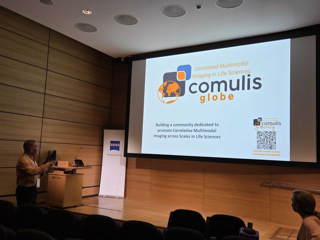
Paul Verkade highlights the COMULISglobe initiative at the 3D CLEM 2026.
The session on “Bridging imaging communities to drive correlative and multimodal imaging” led by Paul Verkade, focused on the need to bring together expertise across different imaging modalities and biological disciplines, highlighting both the vEM and COMULISglobe initiatives. Discussions emphasised that no single researcher or facility can cover the full spectrum of techniques, and that collaboration is essential to overcome technical bottlenecks and advance the field. Discussion participants shared reflections and strategies on how we can continue to strengthen communities in times of scarce funding and how knowledge sharing across domains can be facilitated.
“It was truly impressive to see the pace of technical developments across 3D and correlative imaging that is happening in the community, including at our Nodes."
-- Johanna Bischof, Head of Bio-Hub Operations, Euro-BioImaging
Equally impactful was the panel on “How to make CLEM accessible”, led by Ana Laura Vinagre Costa e Sousa. This discussion reinforced a key message echoed throughout the meeting: that creativity, resourcefulness, and knowledge sharing can make advanced imaging approaches more widely available, even in settings without access to high-end instrumentation.
These panel discussions - introduced as a new format for the meeting - proved to be highly valuable, enabling participants to openly exchange experiences, identify shared challenges, and collectively explore future directions.
Growing momentum in ultrastructural imaging of human tissues
Across multiple presentations and discussions, an important emerging theme was the dynamic developing community around applications of electron microscopy for ultrastructural imaging of human tissues, particularly in a pathology context. Discussions highlighted both the scientific opportunities and the need for coordinated community efforts to develop robust workflows, standards, and access pathways in this area.
This growing interest reflects a broader trend towards integrating high-resolution imaging into biomedical and clinical research, where correlative and multimodal approaches can provide unique insights into disease mechanisms.
A collaborative meeting environment
The success of the symposium was underpinned by excellent organisation and strong support from Zeiss, whose contributions helped create an environment conducive to both scientific exchange and community building.
Overall, the 3D Light and Electron Microscopy meeting demonstrated the strength of the imaging community when working together across modalities and disciplines. For Euro-BioImaging, it highlighted how its Nodes continue to play a central role in driving innovation, enabling access to advanced technologies, and fostering collaboration across Europe in biological and biomedical imaging.
More news from Euro-BioImaging
April 7, 2026
canSERV User Meeting highlights impact and future perspectives for cancer research in Europe
The canSERV Annual Meeting, held in Brussels from 25–27 March 2026, brought together researchers, service providers, Research Infrastructures, policymakers, and patient representatives to reflect…
We were delighted to take part in the 21st European Molecular Imaging Meeting (EMIM), held in Ljubljana, Slovenia, from March 24–27, 2026. The conference…
Euro-BioImaging Welcomes its 2026 Scientific Ambassadors Cohort
Following the continued success of the Scientific Ambassadors programme, Euro-BioImaging is delighted to welcome its 2026 cohort. Building on the strong foundation laid by…