July 1, 2026
Introducing the Euro-BioImaging Annual Report 2025
The Euro-BioImaging Annual Report 2025 is now online! It shares our highlights from 2025, from expanding our community to strengthening our impact, and…

Want to analyze and annotate your sample with the Allen Brain Atlas but don’t have the hardware, software or know-how to do so? Meet Michaela Blazikova, Image Data Analyst at the Institute of Molecular Genetics of the Czech Academy of Sciences. Her facility is part of Euro-BioImaging’s Advanced Light & Electron Microscopy Node in Prague, and they offer Image Data Analysis as a service as part of Euro-BioImaging’s Proof-of-Concept study. In this short article, she shares a compelling example of what Image Data Analysis as a service means.
Michaela Blazikova: In September 2022, we were approached by a user from the Max Planck Institute in Germany who was working on mice stress and behavior analysis. This user had a nice collection of mouse brain images from their home institute (acquired with a light-sheet microscope, Ultramicroscope II from MIltenyi Biotech) that they wanted to share on the Allen Brain Atlas. When the user contacted us, the acquisition and pre-processing had already been accomplished: the clearing of mice brains and imaging was done at MPI and the data were already stitched and fused.
The problem was that for final analysis and annotation, they planned to use an already published analysis pipeline based on specific tools, know-how and hardware they didn’t have ready access to. Specifically, they wanted to use ClearMap, which is a great toolbox for the analysis and registration of volumetric data from cleared tissues, capable of mapping brain activity at capillary resolution. However, it can only be installed on a Linux computer with sufficient performance, and the user didn’t have one. In addition, the workflow requires Jupyter Notebook, an open-source web application for creating and sharing code – but requires knowledge of computer programming languages. In addition, they required some statistical analysis that could be done with MATLAB, but again it requires specialized know-how. Basically, they needed help to finalize their project.
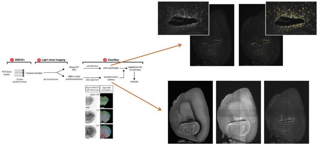
The first step after discussing the project with the user was to check that we had the required hardware and software at our facility. Then, I tested the set up on some test data we had available. In February 2023, once I was sure I could support the user, we agreed on the final output as well as the price. Then we signed the contract.
Currently, I am analyzing the data. It’s our first image data analysis as a service project with Euro-BioImaging, and to carry it out, I’m using one of our brand new remote desktops. It’s impossible to work with such big data on a regular desktop. This project is extremely interesting to me. Furthermore, I can imagine that other users will be potentially interested in this workflow.
So, if you need help with your image data analysis, don’t hesitate to contact us. Maybe we have the tools and expertise to help you out.
About the Advanced Light & Electron Microscopy Prague Node
The Czech Advanced Light and Electron Microscopy Multi Modal Multi Sited Node located in Prague and in České Budějovice offers a wide range of state-of-the-art light and electron microscopy equipment and techniques, offered by five closely collaborating core facilities. The Node is also active in organizing training and courses focused on theoretical and practical aspects of basic and advanced microscopy techniques. Light microscopy instrumentation includes microscopy systems that range from multi-functional point and spinning disc confocal microscopes, light-sheet system up to high-end multi-photon and super-resolution microscopy systems. In the area of electron microscopy, the Node provides expertise and cutting edge equipment for a broad range of biological sample preparation and ultrastructural imaging techniques, including some cryo techniques, electron tomography, SBF-SEM, FIB-SEM and CLEM. Broad expertise in experiment design and data acquisition is also complemented by extensive data analysis services.
July 1, 2026
The Euro-BioImaging Annual Report 2025 is now online! It shares our highlights from 2025, from expanding our community to strengthening our impact, and…

June 26, 2026
Euro-BioImaging, represented by Antje Keppler, Director of the Euro-BioImaging ERIC Bio-Hub, was pleased to contribute to the Irish Universities Association’s Future of Ireland event in…
June 26, 2026
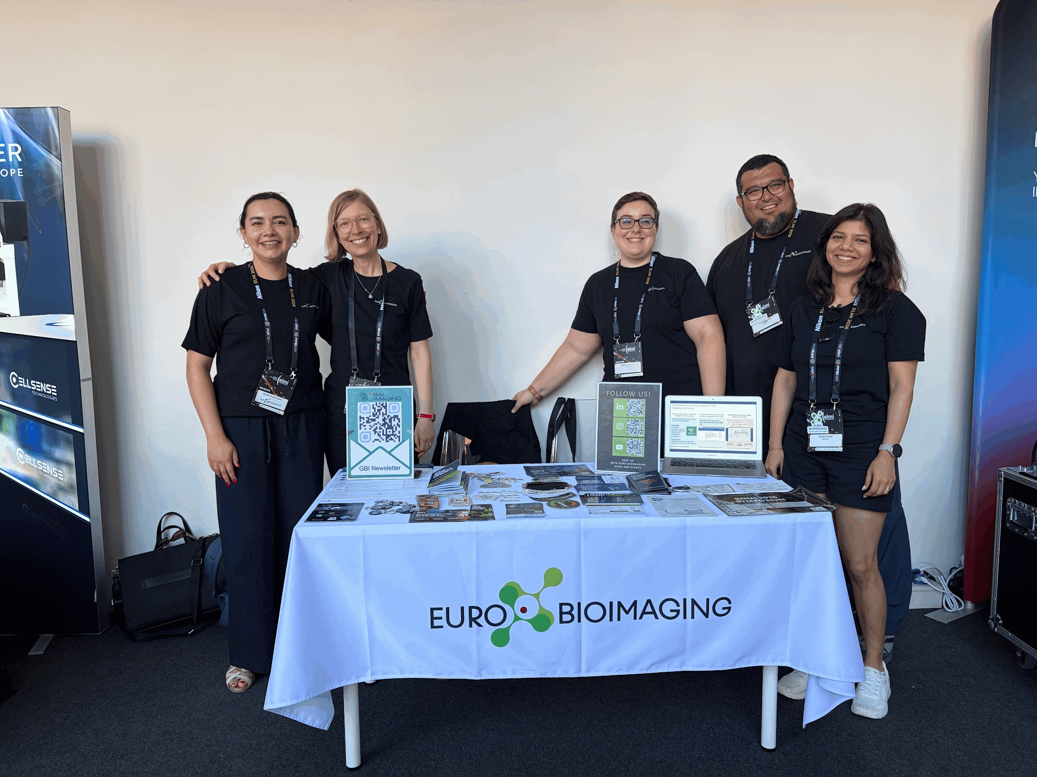
The annual meeting of the European Light Microscopy Initiative (ELMI) took place in Coimbra, Portugal, from June 16-19th, 2026. With nearly 600 in…