Could you tell us a little about yourself briefly?
My name is Dalibor Pánek. I obtained my Ph.D. in applied physics. A year ago I joined the Imaging Methods Core Facility at BIOCEV, part of the Prague node, as a specialist in optical microscopy. I provide training and support for conventional techniques (confocal, TIRF, wide-field epifluorescence) as well as advanced fluorescence microscopy methods (FLIM, multi-photon, imaging FCS, FRET).
We’re talking today about CARS microscopy. Can you briefly tell us what CARS microscopy is and what one can do with it?
CARS (Coherent anti-Stokes Raman scattering) microscopy utilizes nonlinear interactions of two laser beams to image structures based on their vibrational properties, i.e., the contrast is provided by the chemical composition of the observed structure. In principle, chemical mapping of the sample is possible via tunning the two laser beams to various resonant frequencies. Our own system, however, can be used only to detect CARS signal from vibrations of -CH2 residues, which enables imaging of structures with high lipid content. CARS microscopy does not require fluorescence labeling or any special treatment of the sample and can therefore be used on live samples. The resolution of CARS microscopy is about the same as for 2-photon microscopy, so sub-cellular structures can be imaged.
Tell us a bit more about a specific project that was done in your facility using CARS. What scientific questions were you addressing?
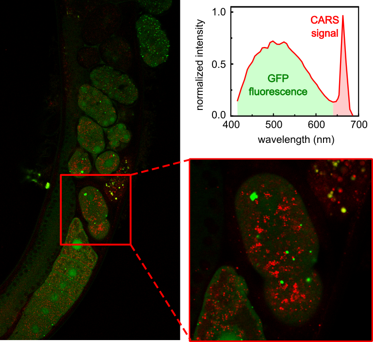
The CARS system in our facility is used for example by the Molecular Pathology research group lead by Z. Kostrouch from the 1st Medical Faculty of the Charles University. In their previous work, they used CARS to visualize lipid structures in C. elegans to study the effect of a particular nematode protein on lipid metabolism, see for example Chughtai et al. 2015 (https://doi.org/10.7717/peerj.1213). They continue this research using the CARS set-up in our facility. However, our data were not published yet. But we think this intriguing technique could be really attractive to new users through Euro-BioImaging.
Simultaneous imaging of GFP-labelled protein, probably responsible for lipid metabolism in C.elegans (green) and unstained lipid droplets by CARS (red) in a living embryo of C. elegans. Data acquired in collaboration with Z. Kostrouch (1st Medical Faculty, Charles University)
Why is CARS best suited to observe lipid structures?
Specific fluorescent labeling of lipid droplets is problematic. Labeling itself can affect the structure and function of the lipids. Therefore, lipid visualization by fluorescence microscopy can yield inconclusive results. CARS does not require labelling of the lipids and therefore provides a reliable way to image lipid-containing structures. Of course, combining CARS microscopy with other complementary imaging techniques is the most advisable approach, as it allows you to see the lipid-containing structures using CARS in relation to other proteins or structures of interest, which can be labelled using fluorescent tags
What are some challenges of CARS? What do researchers have to pay attention to when performing these experiments?
CARS microscopy is a laser scanning technique. Therefore, the user must be prepared to wait a bit to acquire a nice image and importantly while CARS works well for living samples, those samples must be relatively immobile. The CARS imaging set-up is also rather complex. The alignment procedure as well as operation are not straight-forward. But our team is highly experienced, and we always assist users with set-up and alignment.
What other services do you provide in your facility that would be useful in combination with this type of microscopy?
We implemented the CARS modality to extend further the capabilities of already quite powerful laser scanning microscope with a choice of spectral and time-resolved detection. Therefore, multiple techniques can be readily combined in a single multi-modal set-up. Potential techniques that can be combined are conventional fluorescence confocal imaging, 2-photon imaging, second-harmonic generation, and 2-photon-excited fluorescence lifetime imaging (FLIM).
Why should scientists choose your facility for using this technology?
CARS microscopy is a highly specialized technique, commercial systems are quite expensive and as such this technology is not widely available, and rarely offered as an open-access service. A special motivation to use our facility for CARS is the option of the multi-modal acquisitions – allowing combination of CARS with many other imaging modalities to get a full picture of the cellular context. And, of course, scientists can plan their complex experiments not only for a CARS approach but can consider any other methods that are part of the rich portfolio offered in our facility.
How to apply:
In July 2020, Euro-BioImaging launched a Proof-of-Concept study - in collaboration with its Nodes - making it possible to access six imaging technologies previously unavailable via our network. This article – the first in a series – features interviews with Euro-BioImaging Node staff, shedding light on some potential applications of these technologies - and providing advice on how to get the most out of them. We are currently accepting applications to use these technologies as part of the Proof-of-Concept study. Be part of this study - and contribute to community-wide continuous technological innovation!
All scientists, regardless of their affiliation, area of expertise or field of activity can benefit from Euro-BioImaging’s pan-European open access services.Potential users of these new technologies are encouraged to submit project proposals via our website. To do so, you can Login to access our application platform, choose the technology you want to use and the facility you wish to visit, then submit your proposal. All applications will be processed by the Euro-BioImaging Hub. As usual, users will benefit from advice and guidance by technical experts working at the Nodes, training opportunities, and data management services.
Global BioImaging Advance the Conversation on Sustainable Biomedical Imaging Facilities
How can biomedical imaging facilities remain scientifically excellent while becoming more financially, environmentally, and operationally sustainable? These questions were at the heart of…
Moara Lemos: New insights from Global BioImaging’s Job Shadowing programme
Moara Lemos is a Brazilian researcher and imaging scientist. Her award winning research pushes the boundaries of structural biology, using advanced cryo-electron microscopy…
A Multiplexed Vision: Highlights from the Special Edition Virtual Pub on Spatial Transcriptomics
On June 12th, Euro-BioImaging hosted its latest Special Edition Virtual Pub, bringing together over 200 researchers and technology developers working at the cutting…