We are delighted to announce the winners of the “Summer” round of our Four Seasons of the Invisible Imaging Contest. Launched in early 2025, the competition highlights seasonally inspired imaging work that reveals the unseen beauty of the microscopic world. For “Summer,” we were treated to images capturing the season’s warmth and energy, from blooming flowers and intricate seeds to hidden details of tiny parasitic organisms and even a sunrise-like view of a retinal organoid.
We received a high number of creative entries, and our panel of six judges had the difficult task of selecting two winners based on both artistic quality and relevance to the seasonal theme. It was a close call, with just one point between first and second place.
The first prize goes to Tomáš Figuraof Charles University for the image A Sea Brimming with Starfish. The runner-up is Kirstin Vondersteinof Finnish Advanced Microscopy Node, University of Helsinki, for the image Summer’s Hidden Glow: Autofluorescence of Cosmos Bipinnatus.
Tomáš will receive reimbursement of up to €1,000 to attend the scientific conference of his choice, and Kirstin Vonderstein will receive reimbursement of up to €500 to attend a scientific conference.
Check out the breathtaking winning images below and do not miss your chance to take part in the next round of Four Seasons of the Invisible imaging contest! The “Autumn” round is now open for submissions until December 20.
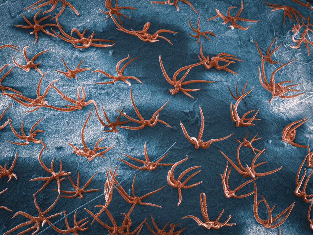
Seed surface of Alyssum Alyssoides by Tomáš Figura
A Sea Brimming with Starfish, by Tomáš Figura, Faculty of Science, Charles University, Prague, Czechia
Resembling a perfect summer holiday scene with a blue sea full of starfish, this is actually the seed surface of Alyssum alyssoides, a xerophilic European plant that produces seeds in the summer. The orange “starfish” structures are trichomes, shadows of “clouds” are likely caused due to insufficient gold coating of the sample. It is scanning electron microscopy image of gold-coated dry sample acquired with a secondary electron detector and subsequently colorized. The author acknowledges technical support from Miroslav Hyliš.
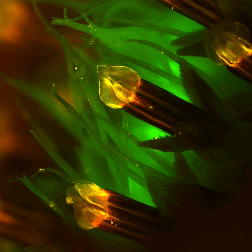
Autofluorescence of Cosmos Bipinnatus, by Kirstin Vonderstein
Summer’s Hidden Glow: Autofluorescence of Cosmos Bipinnatus, by Kirstin Vonderstein, Finnish Advanced Microscopy Node (FiAM), University of Helsinki, Finland
This image captures the stamens of a Cosmos bipinnatus flower that was found blooming along the wayside during the height of summer. Stereomicroscope image (1.63× magnification) of an unstained Cosmos bipinnatus flower, showing natural autofluorescence in yellow, red, and green. Imaging was performed at the Biomedicum Imaging Unit, the University of Helsinki, supported by the Helsinki Institute of Life Science (HiLIFE) and Biocenter Finland.
More news from Euro-BioImaging
July 21, 2026
Building Skills Across Europe: EVOLVE Supports the Second Edition of the Distributed Image Analysis Training Course
The second edition of the “Introduction to Image Analysis with Python for Life Scientists” course marked another successful milestone in the development of Euro-BioImaging’s distributed training model.
Strengthening research-industry partnerships: results from the EVOLVE industry job shadowing pilot
The EVOLVE project, supported by European Union funding, recently completed a pilot programme designed to deepen the relationships between academic research infrastructures…
Imaging-Driven Neuroscience, Responsible Research, and the Power of Advocacy: Highlights from FENS Forum 2026
Euro-BioImaging was pleased to participate in the FENS Forum 2026, Europe’s largest international gathering dedicated to advancing neuroscience (>8000 participants). Throughout…