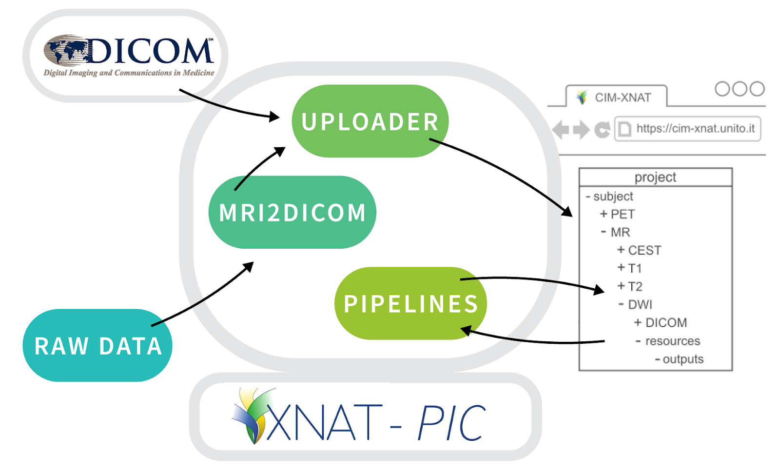
The preclinical imaging community faces many challenges when it comes to collecting, processing, and reusing the various types of imaging data produced in preclinical research. That is why a group from the Molecular Imaging Center (CIM) of the University of Torino, part of Euro-BioImaging’s Multi-Modal Molecular Imaging Italian Node, with support from the Horizon Europe EOSC-Life project, undertook the development of a user-friendly, customizable workflow to store, process and share preclinical image datasets through an XNAT-based platform available at http://cim-xnat.unito.it.
These developments, along with analysis tools for automated image processing, will make it easier for preclinical imaging facilities to manage and process large, multi-modal imaging studies on an XNAT-based system. Depositing these datasets and making them available will promote data sharing among researchers and the reuse of imaging data, and thereby also reducing the need for new animal studies.
The European Open Science Cloud (EOSC) is a digital platform for the European science community, designed to give researchers from the European Union (EU) a global lead in research data management. Within this overarching project, EOSC-Life brings together the 13 Life Science Research Infrastructures (LS RIs) to create an open, digital and collaborative space for biological and medical research. This demonstrator project is an example of how EOSC-Life supports FAIR data principles, as applies to biomedical imaging. EOSC-Life receives funding from the European Union’s Horizon 2020 research and innovation programme under grant agreement No 824087.
More news from Euro-BioImaging
April 9, 2026
Major EU funding for user access & AI development, staff training, data stewardship & many more exciting new services!
Euro-BioImaging ERIC is deeply grateful to announce that the European Union has entrusted our infrastructure with funding to shape the future of imaging and…
Euro-BioImaging is looking for an Operations Support Assistant at the Euro-BioImaging Statutory Seat in Turku, Finland, to support the day-to-day financial and administrative operations…