June 3, 2026
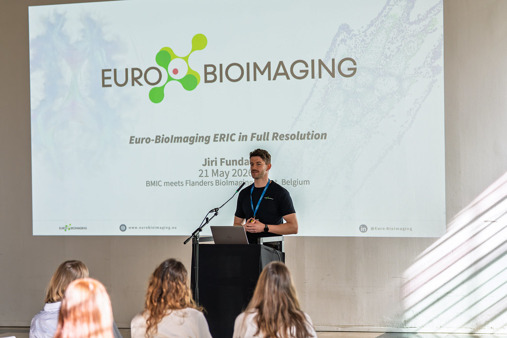
BMIC meets Flanders BioImaging
In May 2026, the Belgian Molecular Imaging Community (BMIC) and Flanders BioImaging joined forces in Ghent for a two-day community event that brought…

We are happy to announce that showcasing of Electron Paramagnetic Resonance (EPR) Imaging is now launched. Used in biomedical research, EPR imaging is a non-invasive, in vivo methodology used to determine tissue microenvironment parameters. Euro-Bioimaging Nodes are invited to participate in a showcase to make this technology available in open access.
To support the imaging community and its associated high-quality research, Euro-BioImaging must remain at the technological forefront. Thus, a workflow has been established, to ensure that new technologies are continuously integrated by the community into the Euro-BioImaging portfolio.
Showcasing is the first step in this workflow. It is aimed at demonstrating:
a) User need and relevance for the scientific community (through e.g. documentation by technology hosts on successful external user access to this technology; submitted letters of interest from users; relevant research publications associated with this technology in general, etc.)
b) An operational access model for external users to this technology (through documentation of access model by technology hosts).
A showcase can be conducted by any technology developer/provider or imaging facility at a public research institution/university in Europe, which is offering the new technology and hosts external users to apply this technology in their research. For more information please visit this page.
If you wish to showcase EPR Imaging, please let us know by sending an email at info@eurobioimaging.eu . We’ll be happy to support you with the process!
June 3, 2026
In May 2026, the Belgian Molecular Imaging Community (BMIC) and Flanders BioImaging joined forces in Ghent for a two-day community event that brought…

June 1, 2026
The EOSC Association has adopted a new position paper calling for a distinct Work Programme-based Partnership for EOSC in FP10. Euro-BioImaging welcomes this…
May 29, 2026
Spatial transcriptomics is rapidly transforming the way researchers study complex biological systems. To explore the latest developments in this fast-moving field, Euro-BioImaging is…