In May 2025, Anil Can from Acıbadem Üniversitesi in Istanbul traveled to Prague to explore the fine structure of mitochondria in testicular tissue. Supported by the Imaging4All (i4A) programme, coordinated by Global BioImaging and funded by the Wellcome Trust, Anil spent two weeks at the Electron Microscopy Core Facility (IMG EM) of the Institute of Molecular Genetics of the Czech Academy of Sciences, part of the Advanced Light and Electron Microscopy Prague Node of Euro-BioImaging.
His project focused on studying mitochondrial damage caused by MSG and the ameliorating effects of ferulic acid on this—a topic with important implications for understanding toxicity impacts in male reproductive health. The entire IMG EM team, including Facility Lead Vlada Filimonenko and EM specialist Dominik Pinkas, supported the project using Focused Ion Beam Scanning Electron Microscopy (FIB-SEM) and Transmission Electron Microscopy (TEM) tomography to capture the ultrastructure of mitochondria in three dimensions. We talked to Vlada and Dominik to get their view on the experience of hosting this user project.
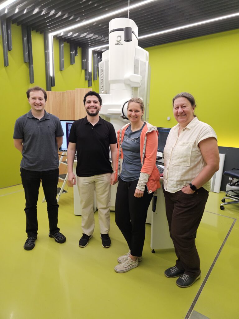
Dominik Pinkas, Anil Can, Marketa Dalecka, and Vlada Filimonenko in front of the Electron Microscope.
Adapting to challenging samples
Anil arrived with already fixed and resin-embedded mouse testicular tissue, prepared using protocols different from those typically used at the facility. “Normally, we fine-tune the sample preparation here to match the intended imaging method,” explains Vlada. “So we weren’t entirely sure how well these samples would perform.”
To accommodate the material, the team adjusted their imaging workflow and optimized acquisition parameters to deal with lower sample contrast. “The two imaging methods used usually require a slightly different sample preparation approach, it’s a careful balance between structure preservation, contrast and resolution,” says Dominik, “but the results were very good in the end.”
Hands-on collaboration and training
During his stay, Anil was actively involved in every step of the project—from selecting regions of interest to image acquisition and data reconstruction. “He had no previous experience operating electron microscopes and the vEM methods we used for the project are quite complex, so there was a huge amount to learn,” recalls Vlada. “But he was very engaged, and his input was crucial for identifying the right areas to image.”
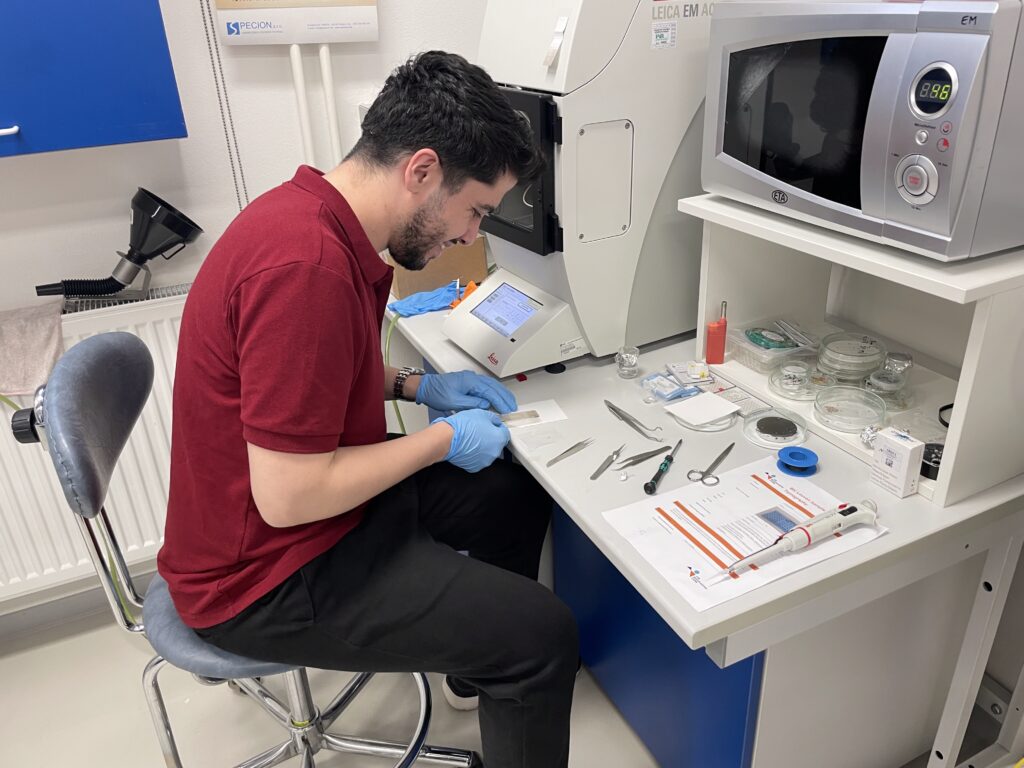
Anil Can during the sample preparation stage.
The facility provided intensive training on both data acquisition and processing, ensuring that Anil could continue working independently with the large datasets once back home. Data transfer was facilitated through the Czech CESNET network, and the facility continues to provide advice as Anil analyses the large quantities of gathered data.
Reflecting on the experience, Anil shared:
“During my stay, I gained first-time hands-on experience with TEM tomography and FIB-SEM, and we obtained high-quality 3D micrographs from testicular tissue samples. Stepping into the volumetric world of electron microscopy was truly fascinating—each 3D dataset felt like uncovering an entirely new layer of biological reality.”
To allow the i4A project to go ahead does not just require support from the host institution, but also from the grantees home institution. Anil stresses that “I am grateful to my advisor, Dr. Merve Açıkel Elmas, whose scientific guidance and continuous support have shaped both this project and my development as a researcher.”
A rewarding change of pace
For the IMG EM team, hosting an Imaging4All project meant focusing fully on one international collaboration for two weeks—a change from their usual multi-project schedule.
“It’s quite intense but also refreshing,” says Dominik. “We give our full attention to every user project, but in this case, everything happened in a concentrated period. It’s rewarding to see the results develop so quickly.”
Early communication was key to success. The facility and Anil discussed the experiment months in advance, revising the approach and selecting the most suitable imaging methods before the application was submitted. This preparation, alongside well-organised scheduling of the entire facility team, ensured that the visit itself ran efficiently and that both instrument time and user training were maximized.
Strengthening open-access science
The project gave Anil access to advanced methods that are otherwise unavailable in his home institution, while also expanding the facility’s international network. “The scientific quality of i4A projects is very high thanks to their rigorous evaluation,” says Vlada. “And since they are fully funded, we can focus entirely on the science and on supporting the visiting researcher.”
Looking ahead, the IMG EM Core Facility team is eager to host future Imaging4All users. “We’re already talking to new potential applicants,” says Vlada. “These projects are a great way to connect expertise across borders and to show how open-access infrastructures like Euro-BioImaging can make a real difference.”
About Imaging4All
Imaging4All (i4A) is a mobility funding programme that enables researchers from low- and middle-income countries to access imaging facilities anywhere in the world. Coordinated by Global BioImaging and funded by the Wellcome Trust, the programme supports scientific exchange, training, and global collaboration through open-access imaging science.
More news from Euro-BioImaging
April 9, 2026
Major EU funding for user access & AI development, staff training, data stewardship & many more exciting new services!
Euro-BioImaging ERIC is deeply grateful to announce that the European Union has entrusted our infrastructure with funding to shape the future of imaging and…
Euro-BioImaging is looking for an Operations Support Assistant at the Euro-BioImaging Statutory Seat in Turku, Finland, to support the day-to-day financial and administrative operations…