
March 26, 2026
Welcome, MAdMiC Node!
The Madrid Advanced Microscopy Center (MAdMiC) is the first Euro-BioImaging Node in Madrid (Spain). It is formed through the collaboration and close work of…

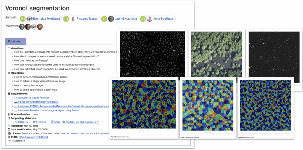
The first major outcome of the OSCARS FIESTA project is a brand new Galaxy training tutorial on Voronoi-based segmentation. Developed from a cross-disciplinary perspective, the tutorial introduces the workflow using tools that are freely available, open source, and designed to work across research domains.
Whether you're analysing microscopy images of cells, astronomical data from deep space, or environmental samples, Voronoi segmentation offers an intuitive and effective approach for separating densely packed objects without the need for AI or pre-trained models.
The tutorial walks users through a step-by-step workflow in Galaxy, starting from a raw image and producing a clean segmentation using Voronoi diagrams. This classic technique partitions space based on object centers, enabling researchers to separate tightly grouped instances with minimal preprocessing.
The result is a robust, FAIR workflow that works equally well on images of cell colonies, droplets, or other clustered biological structures.
The OSCARS FIESTA project — funded by the OSCARS project — is working to break down disciplinary silos in image analysis by fostering reusable, cross-domain workflows. By connecting bioimaging, astrophysics, and environmental sciences, the project aims to build a truly FAIR ecosystem for scientific image data.
The tutorial is part of the Galaxy Training Network (GTN), ensuring it’s fully open and ready to be reused or adapted by trainers and researchers alike.
This tutorial is just the beginning. Stay tuned for more news from OSCARS FIESTA — and in the meantime, try out the tutorial and share your feedback!
Access the tutorial: https://training.galaxyproject.org/training-material/topics/imaging/tutorials/voronoi-segmentation/tutorial.html
March 26, 2026
The Madrid Advanced Microscopy Center (MAdMiC) is the first Euro-BioImaging Node in Madrid (Spain). It is formed through the collaboration and close work of…
March 26, 2026
German BioImaging, within its work in the NFDI4BIOIMAGE consortium, and in collaboration with Euro-BioImaging ERIC, has launched a new survey to collect input about…
March 25, 2026
Turin, Italy – 20–22 October 2026 Early career professionals working in imaging core facilities will soon have the opportunity to strengthen essential skills beyond…