Through the canSERV project, Euro-BioImaging Nodes continue to support innovative cancer research across Europe. One recently completed user project, carried out at the University Medical Center in Utrecht, part of our Dutch High Field MR Node, has demonstrated for the first time the feasibility of performing Deuterium Metabolic Imaging (DMI) in the human breast.
The project originated after Dr. Claudius Sebastian Mathy, from the Friedrich-Alexander University Erlangen-Nuremberg, learned about canSERV from prof. Dennis Klomp, Head of the Dutch High Field Imaging Hub Node, during the ISMRM annual meeting in Singapore in 2024 and submitted a proposal to the programme’s second open call. Following approval in November 2024, preparations were completed and the proof-of-concept study was conducted at Utrecht UMC between March and September 2025.
Exploring a new metabolic imaging approach for breast cancer
DMI combines 2H Magnetic Resonance Spectroscopic Imaging (MRSI) with the administration of deuterium-labelled substrates, enabling the direct detection of metabolic processes in vivo.
Since breast cancer remains the most common cancer in women, researchers are actively exploring methods that can reveal tumour metabolism early and more specifically, potentially improving treatment response assessment.
Until now, DMI applications in breast cancer research were limited to animal models. This project marked the first time DMI has been applied to the human breast.
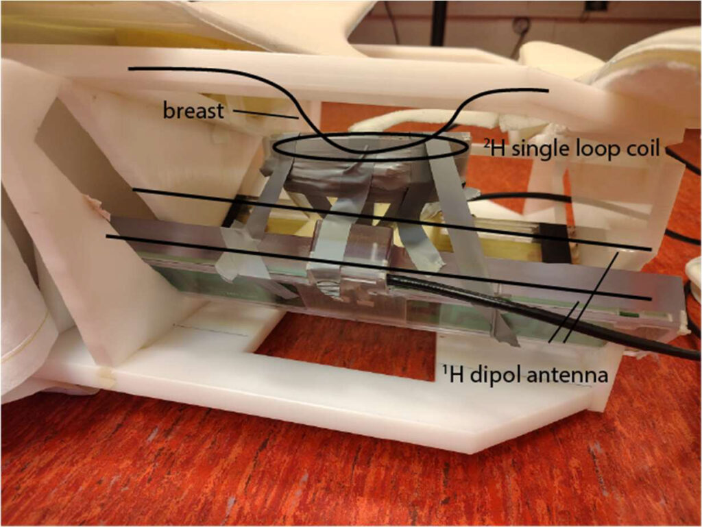
Developing the measurement setup
Working closely with the Utrecht UMC team, the project developed a coil configuration and acquisition workflow for unilateral breast scans at 7T, integrating transmit and receive elements for both 2H and 1H imaging. After phantom testing, a series of feasibility scans were acquired from healthy volunteers both at natural deuterium abundance and following oral administration of 2H-labelled glucose.
Schematic drawing of the setup to detect 2H and 1H MR signals in breast unilaterally in prone position.
“This simply would not have been possible without access to the equipment, expertise, and technical support at Utrecht UMC. The collaborative environment was essential for building and validating the setup.”
-- Dr. Claudius Sebastian Mathy, from the Friedrich-Alexander University Erlangen-Nuremberg
Feasibility demonstrated in healthy volunteers
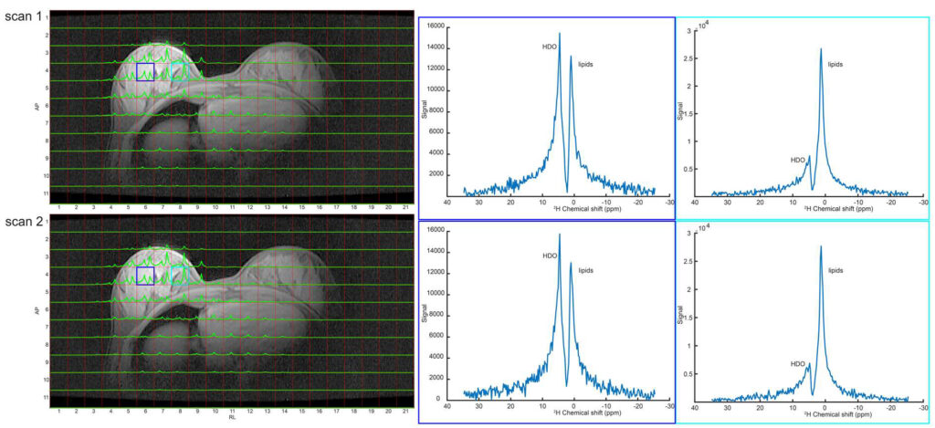
The acquired data demonstrated reproducibility of natural-abundance 2H signals and metabolic response following substrate administration, indicating that DMI can detect metabolism in human breast tissue. The study provides a technical foundation for future patient studies.
“We were able to show that not only is DMI in the breast technically feasible, but the metabolic signals are stable and quantifiable. This is a crucial step toward clinical translation.”
-- Dr. Claudius Sebastian Mathy, from the Friedrich-Alexander University Erlangen-Nuremberg
Results have already been presented at international conferences, and further dissemination is planned.
Axial slice of two natural abundant 2H MRSI scans acquired after each other of a healthy volunteer (2H receiver loop beneath the right breast). Selected voxels (indicated in blue and red) containing different ratios of glandular breast tissue and fat shows peaks from HDO, set to 4.7 ppm and a lipid signal at 1.0-1.3 ppm. Almost similar signal intensities of both 2H MRSI scans (submitted to ISMRM).
Looking ahead
The project highlights the impact of canSERV funding in enabling cutting-edge biomedical imaging studies by accelerating access to highly specialised research infrastructures.
“This preliminary work gives us a strong foundation to apply for future funding. Our long-term vision is to evaluate whether DMI can contribute to earlier and more precise treatment monitoring in breast cancer patients.”
-- Dr. Claudius Sebastian Mathy, from the Friedrich-Alexander University Erlangen-Nuremberg
More news from Euro-BioImaging
July 23, 2026
Cellular Imaging Hungary Node expands expertise in advanced neurophotonics
The Euro-BioImaging Cellular Imaging Hungary Node has expanded its service portfolio with the addition of the BrainVisionCenter (BVC) in Budapest. Following a successful…
The UK Euro-BioImaging Node expands from seven to thirteen sites!
We’re delighted to announce that the UK Euro-BioImaging Node is expanding, growing from seven to thirteen sites following a successful upgrade application and…
Building Skills Across Europe: EVOLVE Supports the Second Edition of the Distributed Image Analysis Training Course
The second edition of the “Introduction to Image Analysis with Python for Life Scientists” course marked another successful milestone in the development of Euro-BioImaging’s distributed training model.