How can synchrotron-based imaging move from experimental research towards clinical applications? This was among the questions explored at the International Workshop on Pulmonary Functional Imaging (IWPFI) 2026 in Mont-Tremblant, Canada, where researchers from around the world gathered to discuss advances in pulmonary imaging.
Among the participants were Claudia Victoria Benke, currently Euro-BioImaging Scientific Ambassador, and Prof. Dr. Christian Dullin, former Scientific Ambassador. Both are part of an international research collaboration exploring the potential of synchrotron-based X-ray phase-contrast computed tomography (CT) for in vivo patient lung imaging.
Their recent study, published in The Lancet Medical Imaging & Theranostics, demonstrated the technical feasibility of imaging a fresh, unfixed human lung using high-resolution phase-contrast CT with clinical acceptable x-ray dose. The research was conducted at Euro-BioImaging's Phase Contrast Imaging Flagship Nodeat Elettra Sincrotrone Trieste, Italy, contributing to ongoing efforts to develop this technology for future clinical applications.
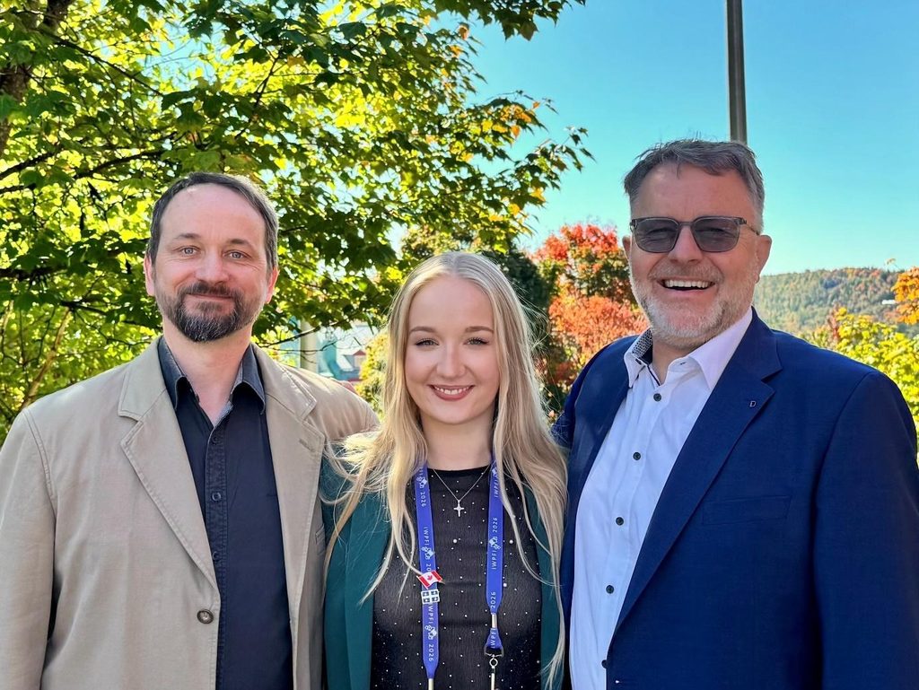
Prof. Dr. Christian Dullin (left), former Euro-BioImaging Scientific Ambassador, Claudia Victoria Benke (centre), current Scientific Ambassador and first author of The Lancet Medical Imaging & Theranostics publication, and Prof. Dr. Hans-Ulrich Kauczor (right) at IWPFI 2026 in Mont-Tremblant, Canada.
Seeing the lung in greater detail
The human lung contains an intricate network of airways, blood vessels and delicate tissue structures. Although conventional CT is an essential diagnostic tool, its spatial resolution limits the visualisation of the lung's finest anatomical features.
Synchrotron phase-contrast CT offers a different approach. Rather than relying primarily on X-ray absorption, it exploits subtle changes in the phase of X-rays passing through tissue, enhancing contrast between structures that are otherwise difficult to distinguish.
To investigate its capabilities, the research team examined a fresh, unfixed human lung obtained during forensic autopsy at the University of Trieste, in collaboration with Prof. Dr. Stefano D’Errico, head of Forensic Medicine at the university. The specimen was positioned inside a specially designed anthropomorphic chest phantom to maintain lung inflation during imaging at Elettra's SYRMEP beamline.
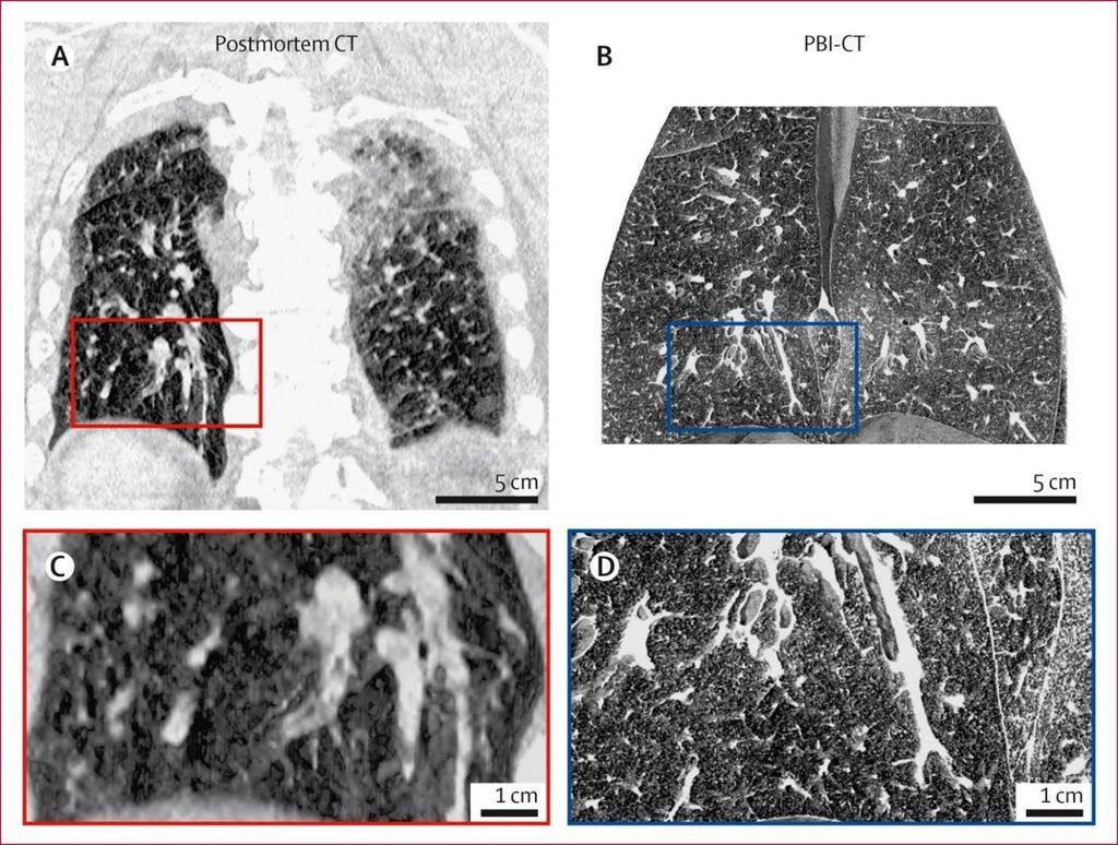
The resulting images achieved an effective voxel size of 67 micrometres, revealing fine pulmonary structures, including small airways, blood vessels, interlobular septa and secondary pulmonary lobules. Compared with conventional postmortem CT, the phase-contrast images showed greater anatomical detail and less extensive density artefacts, although the examinations were performed under different lung inflation conditions.
The study illustrates how specialised imaging infrastructure, together with expertise in medical imaging, synchrotron science and forensic medicine, enables new approaches to investigating human lung anatomy.
Comparison of conventional postmortem CT and synchrotron phase-contrast CT (PBI-CT) of a human lung. The phase-contrast images demonstrate improved visualisation of fine pulmonary structures, including small airways, vessels and interlobular septa. From Benke et al., The Lancet Medical Imaging & Theranostics (2026), CC BY 4.0.
Bringing synchrotron imaging closer to patients
Beyond its application in forensic imaging, the study contributes to the development of synchrotron phase-contrast CT for potential clinical use.
An important step towards this goal is the planned SYRMEP-LS beamline at Elettra Sincrotrone Trieste. Led by Dr. Giuliana Tromba the facility is anticipated to become operational in 2028 and is intended to investigate the technique in selected patients whose conventional CT examinations have produced inconclusive findings.
The proposed approach would focus on targeted imaging of regions requiring further investigation, potentially providing additional anatomical information rather than replacing conventional CT.
Previous experimental studies have demonstrated the feasibility of synchrotron phase-contrast lung imaging at radiation dose levels comparable to clinical CT. The recent Lancet publication builds on this work by demonstrating whole-organ imaging of fresh human lung tissue. However, further technological development and clinical validation remain essential before imaging living patients.
Sharing research and building connections at IWPFI 2026
At IWPFI, Benke presented the team's work on synchrotron phase-contrast CT and discussed the next steps towards clinical translation. Dullin presented complementary research on multi-scale and multi-modal imaging of lung tissue.
The conference brought together researchers from across the pulmonary imaging community, providing opportunities to exchange ideas, explore new collaborations and identify shared research interests. These discussions offered fresh perspectives for the team's ongoing work.
Benke's participation was supported by Euro-BioImaging and the Rolf W. Günther Foundation.
Publication
Benke CV, Dullin C, Radaelli D, et al. Synchrotron phase-contrast CT of a human lung from forensic autopsy: a single-case technical feasibility study. The Lancet Medical Imaging & Theranostics. 2026;1:100025. DOI: https://doi.org/10.1016/j.lanmit.2026.100025
This article was written by Scientific Ambassador Claudia Victoria Benke
More news from Euro-BioImaging
November 17, 2026
Dutch Microscopy Days 2026 brings together light and electron microscopy communities
On 2–3 November 2026, around 180 microscopy experts gathered in Maastricht for the second edition of the Dutch Microscopy Days. Jointly organised by…
Euro-BioImaging at EMBL's Festival of Science & Curiosity
On Friday, 25 September 2026, Euro-BioImaging Bio-Hub team members joined the European Researchers’ Night celebrations at EMBL Heidelberg’s Festival of Science and Curiosity.
Building connections for the future of Danish Life Science Research Infrastructures
How can research infrastructures build sustainable futures, strengthen collaboration and support the people who make them work? These questions were at the heart…