Cassava is one of the world’s most important staple crops, providing a primary source of carbohydrates for nearly 500 million people across tropical regions (source: Wikipedia). Despite its resilience, cassava production is threatened by devastating plant viruses such as the ipomovirus responsible for Cassava Brown Streak Disease (CBSD). Preventing major outbreaks is crucial for global food security, particularly in regions where cassava is a daily dietary staple.
To better understand how the Ugandan cassava brown streak virus (UCBSV), one of the causal agents, behaves — and how cassava plants may resist infection — researchers Dr. Samar Sheat from the Department of Plant Viruses at the Leibniz Institute DSMZ in Braunschweig, Germany, and Somruthai Chaowongdee from the Department of Plant Pathology at Kasetsart University in Thailand have joined forces. Somruthai’s research is conducted under the supervision of Dr. Wanwisa Siriwan, a virologist and director of the Hub of Knowledge and Technology Center of Cassava in Thailand, in close collaboration with Dr. Samar Sheat. Supported by Imaging 4 All, a Global BioImaging initiative funded by the Wellcome Trust, the team is using cryo-electron microscopy (cryo-EM) and 3D helical reconstruction at the EMBL Imaging Centre to generate the first high-resolution structural reference of UCBSV virions.
Mapping a virus for the first time
While quarantine measures have helped countries such as Thailand — the world’s largest exporter of cassava starch — to avoid major CBSD outbreaks so far, long-term protection depends on developing virus-resistant cassava varieties. Achieving this requires a detailed understanding of the virus itself, including its structure and replication mechanisms. Until now, however, the structure of the virus responsible for CBSD had never been mapped.
High-resolution electron microscopy is essential for uncovering the structural organisation of viral coat proteins and understanding how the virus interacts with its host and insect vectors. However, access to advanced Electron Microscopy infrastructure remains limited in many regions. In Thailand, for example, only one university currently provides electron microscopy services, and waiting times can be extremely long.
Transformative collaboration opportunity
For Somruthai, access to the imaging technologies at Euro-BioImaging’s EMBL Node through Imaging 4 All was transformative.
“Imaging 4 All really made this research possible,” she says. “This funding allows us to work together and do something that has never been done before.”
The collaboration combines complementary expertise and resources. Samar’s home institution, the Leibniz Institute DSMZ, hosts one of the world’s most comprehensive plant virus collections. For this project, the team chose to work with wild-type UCBSV virions to define coat protein organisation, analyse helical symmetry, and investigate structural features that may influence virus transmission.
Preparing the samples for cryo-EM was a lengthy and meticulous process. After the grant was awarded, Somruthai travelled to Braunschweig to work alongside Samar, and together they spent nearly two months preparing samples for imaging.
Once at the EMBL Imaging Centre, however, the team quickly began generating results with the support of cryo-EM specialist Simon Fromm.
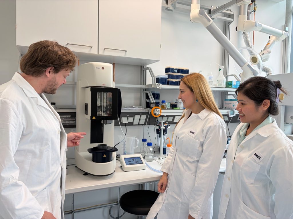
Simon Fromm (left), Samar Sheat (centre) and Somruthai Chaowongdee (right) in the EMBL lab.
Fast preliminary results
“Within the first 24 hours, we could already see the virus,” says Samar. “It’s amazing to observe it with high-resolution microscopy.”
The EMBL Imaging Centre’s full user support model was key to the project’s rapid progress.
“If we can successfully characterise the wild-type virus, we may then be able to study recombinant viruses to investigate vector transmission efficiency and host adaptation. A promising first result is essential for securing future funding and continuing this research.”
-- Samar Sheat, DSMZ Leibniz
“This virus is relatively straightforward to work with if you have experience,” explains Simon. “But teaching new users how to optimise data collection and processing would take much longer. Providing hands-on support allows researchers to focus on generating meaningful results.”
For Simon, working with the ipomovirus for the first time was also exciting.
“It’s a nice sample because it is large enough to be visible directly in the raw microscope images,” he says. “Many samples are much smaller and only appear as grey dots until image processing confirms what you are looking at.”
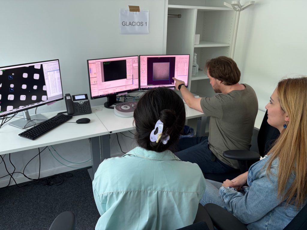
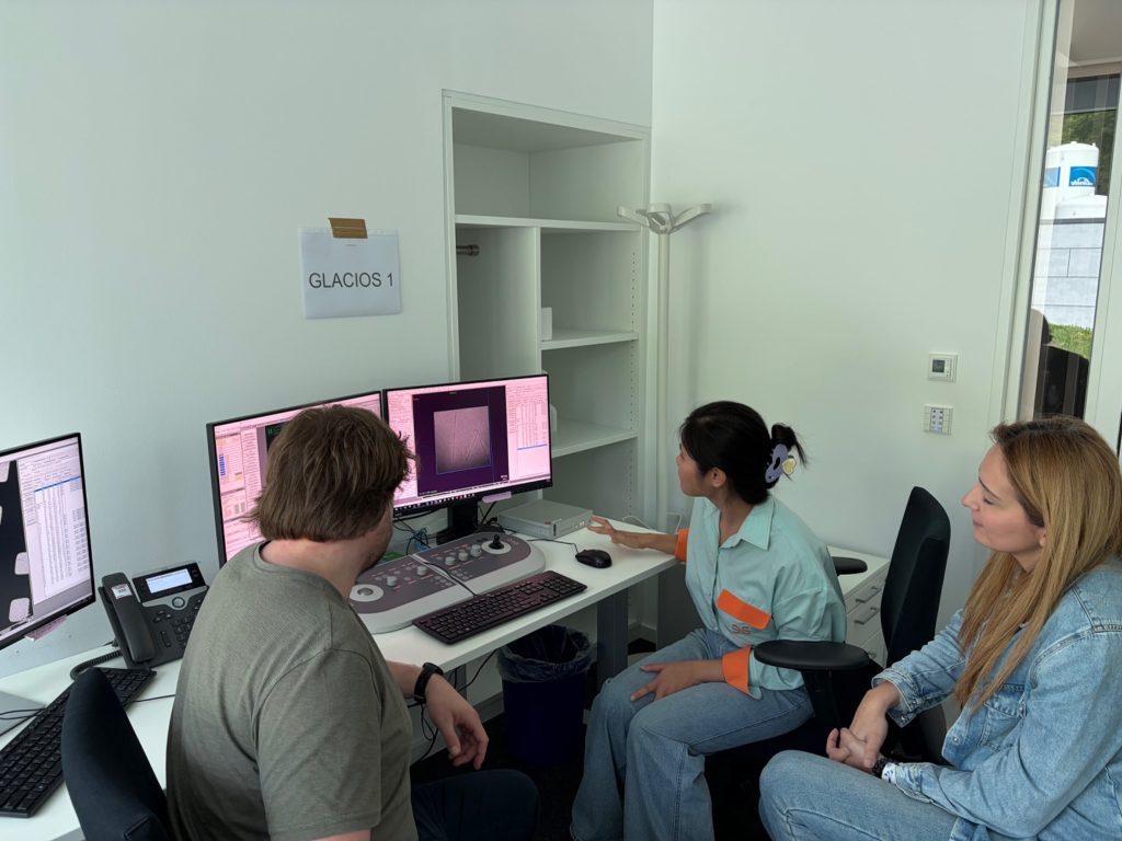
Simon Fromm, Samar Sheat and Somruthai Chaowongdee look closely at the first images from the Electron Microscope.Somruthai looks closely at preliminary images from the Electron Microscope.
A range of new skills
During her stay at EMBL, Somruthai not only collected data but also gained valuable experience in project organisation, sample preparation, and cryo-EM workflows.
“I now better understand how Electron Microscopy works and how to prepare samples more effectively in the future,” she says.
For the researchers, this pilot study represents an important first step toward larger future projects.
“If we can successfully characterise the wild-type virus, we may then be able to study recombinant viruses to investigate vector transmission efficiency and host adaptation ” explains Samar. “A promising first result is essential for securing future funding and continuing this research.”
Potential for future and real-world impact
The project also highlights the broader importance of international collaboration and equitable access to imaging technologies. By connecting researchers from different regions with advanced imaging facilities and expertise, initiatives such as Imaging 4 All help address global scientific challenges that directly impact food security and agriculture. The knowledge generated through this collaboration could ultimately contribute not only to future research funding and policy development, but also to protecting cassava crops and the communities that depend on them.
“Imaging 4 All really made this research possible. This funding allows us to work together and do something that has never been done before.”
Cellular Imaging Hungary Node expands expertise in advanced neurophotonics
The Euro-BioImaging Cellular Imaging Hungary Node has expanded its service portfolio with the addition of the BrainVisionCenter (BVC) in Budapest. Following a successful…
The UK Euro-BioImaging Node expands from seven to thirteen sites!
We’re delighted to announce that the UK Euro-BioImaging Node is expanding, growing from seven to thirteen sites following a successful upgrade application and…
Building Skills Across Europe: EVOLVE Supports the Second Edition of the Distributed Image Analysis Training Course
The second edition of the “Introduction to Image Analysis with Python for Life Scientists” course marked another successful milestone in the development of Euro-BioImaging’s distributed training model.