
March 26, 2026
Welcome, MAdMiC Node!
The Madrid Advanced Microscopy Center (MAdMiC) is the first Euro-BioImaging Node in Madrid (Spain). It is formed through the collaboration and close work of…

Xylella fastidiosa is a vector-borne gram-negative xylem-limited bacterium native to the Americas, affecting more than 650 plant species, including crops of major economic importance. The bacterial sequence type ST53 is the causal agent of the Olive Quick Decline Syndrome, that devastated the Southern Italian olive heritage. The meadow spittlebug Philaenusspumarius (Hemiptera: Aphrophoridae) is the only epidemiologically relevant insect vector identified in all the European bacterium outbreaks so far.
Since the first detection of the bacterium in Europe, research has mostly focused on the bacterium-plant interaction, overlooking aspects of the insect vector-plant interaction which are crucial for the bacterium spread. For example, there is a significant knowledge gap on plant traits underlying host plant location and acceptance by the vector, and that could make a plant more or less attractive for the insect.
Hamouche Zeineb, a PhD student under the supervision of Prof. Daniele Cornara at the University of Bari Aldo Moro - Department of Soil, Plant and Food Sciences (DiSSPA), is working on investigating anatomical and chemical traits in different olive varieties and how they shape spittlebug-olive interaction, eventually shaping bacterial spread.
Such insights are crucial for designing sustainable control strategies that reduce vector-host contact and subsequent pathogen transmission.
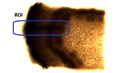
Hamouche Zeineb is a PhD student at the University of Bari Aldo Moro - Department of Soil, Plant and Food Sciences (DiSSPA) who has received funding from the AgroSERV project (No. 101058020) first call for access to use Euro-BioImaging's research facilities to further elucidate spittlebug stylets route and its penetration impact in olive varieties with different susceptibility to Xylella fastidiosa.
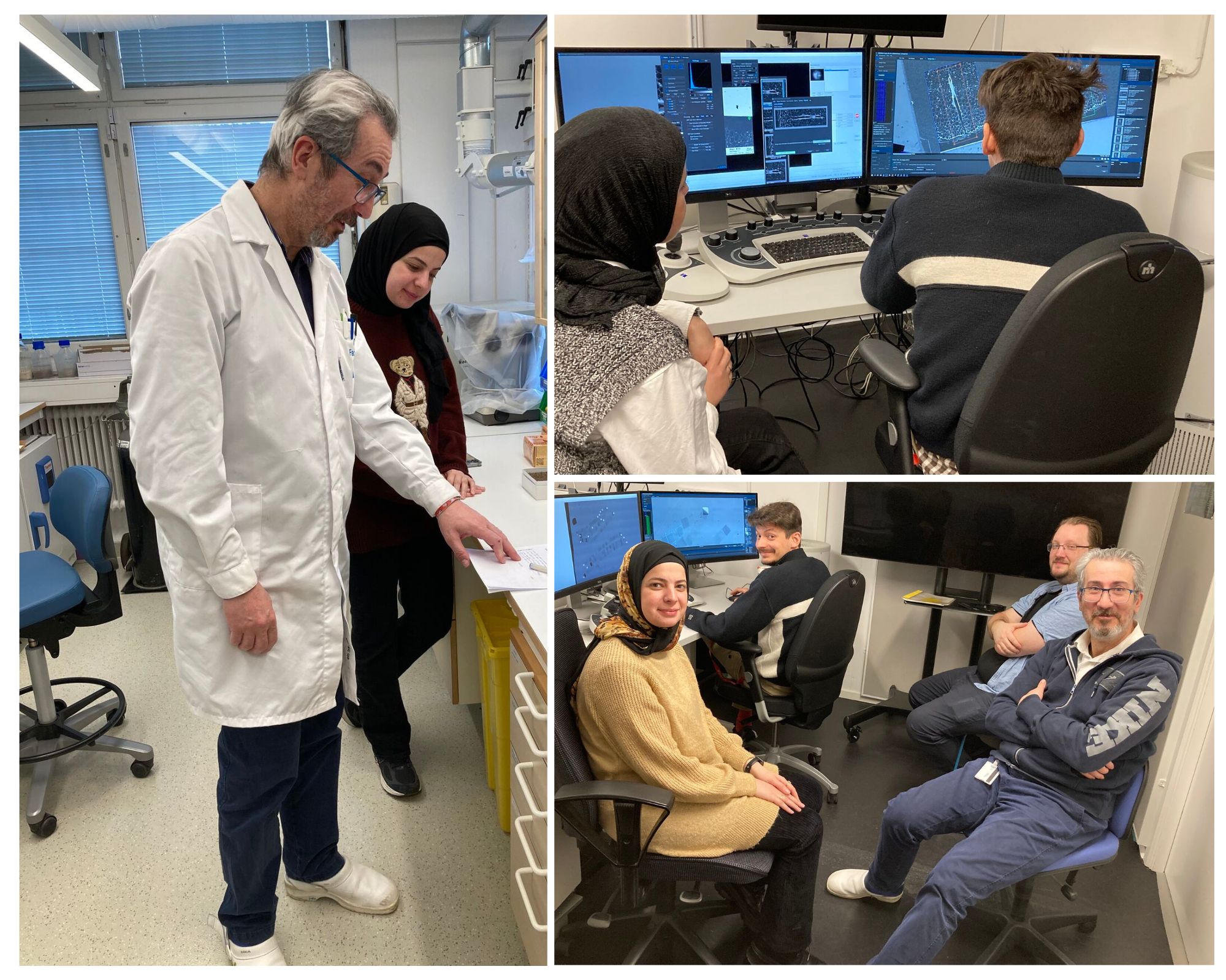
Accessing the Three-Dimensions Volume Scanning Electron Microscopy (Array Tomography) technology provided by the Centre for Cellular Imaging at the University of Gothenburg — a National Microscopy Infrastructure (NMI) Sweden Node and part of the Swedish Euro-BioImaging Node, has allowed Zaineb to shed light on overlooked aspects of the impact of spittlebug stylets penetration inside olive plant tissues and feeding dynamics.
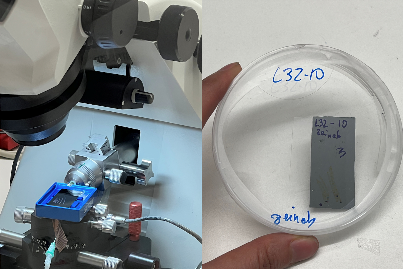
During her visit, Zeinab skillfully navigated the entire imaging pipeline—from sample preparation and sectioning to high-resolution image acquisition, data processing, and analysis—making a significant contribution to knowledge transfer and capacity building.
Cutting-edge imaging technologies, like 3D volume Scanning Electron Microscopy, are opening a new frontier in agricultural science. These powerful tools are giving researchers an unprecedented view into the complex interactions between crop plants and the pests that threaten them.
By uncovering the intricate relationship between the meadow spittlebug and olive plants, researchers are now able to elucidate crucial aspects of the insect vectors-plant interaction underlying X. fastidiosa transmission dynamics and pathogen spread. This leap in understanding represents a vital milestone in the fight to protect olive groves across the Mediterranean, where X. fastidiosa has caused widespread damage. More importantly, it paves the way for smarter, more sustainable pest management strategies—ones that reduce reliance on chemical treatments, safeguard crop yields, and help preserve the delicate balance of biodiversity within agricultural ecosystems
With applications that extend across the agricultural sector, these advanced imaging tools promise to reshape the future of crop protection, offering innovative, and environmentally friendly approaches to safeguarding food security and cultural heritage. Advanced imaging isn’t just changing how we see pests, It’s changing how we secure our food.
This success story highlights the transformative impact of European Research Infrastructures, like Euro-BioImaging, and EU-funded initiatives, such as AgroSERV, in advancing scientific discovery. By providing widespread access to state-of-the-art imaging technologies, Euro-BioImaging empowers researchers to deepen their understanding of plant health and disease, contributing to a more sustainable and resilient food system for all.
Article written by Ayoub El Ghadraoui
March 26, 2026
The Madrid Advanced Microscopy Center (MAdMiC) is the first Euro-BioImaging Node in Madrid (Spain). It is formed through the collaboration and close work of…
March 26, 2026
German BioImaging, within its work in the NFDI4BIOIMAGE consortium, and in collaboration with Euro-BioImaging ERIC, has launched a new survey to collect input about…

March 25, 2026
Turin, Italy – 20–22 October 2026 Early career professionals working in imaging core facilities will soon have the opportunity to strengthen essential skills beyond…