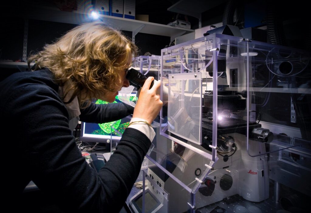
Euro-BioImaging’s French Node in Bordeaux is contributing to an important study led by the University of Bordeaux to understand COVID-19 infection and inflammatory response using fully differentiated human bronchial epithelium as model. Fluorescent imaging techniques such as immune fluorescence and RNAscope technology will be used in this highly relevant physiological system to determine if a particular cell type is (preferentially) infected by the virus. Dr Harald Wodrich, INSERM Research Director at the University of Bordeaux, explains.
The study, called ANACONDA, is funded by the French ANR Flash COVID-19 call. It combines the unique expertise of four local partners: Dr. Thomas Trian, from the Centre de Recherche Cardio-Thoracique de Bordeaux, INSERM U1045, who had previously developed a model of differentiated airway epithelia to study asthma, Prof. Denis Malvy, head of the tropical and infectious diseases unit at the CHU and regional coordinator of the SARS-CoV-2 response, Marie-Line Andreola and Harald Wodrich, from the MFP CNRS UMR 5234 at the University of Bordeaux, experts on highly pathogenic RNA-viruses and virus microscopy, and the Bordeaux Imaging Center for virus imaging at the cellular level.
Interdisciplinary collaboration
The objective of the study is to understand the contribution of the bronchial epithelium to the immune response triggered by SARS-CoV-2 infection and causing high morbidity. SARS-CoV-2 infection will be analysed according to the presence of respiratory diseases such as COPD. Different risk factors (age, gender, tobacco consumption and diabetes) will also be taken into account. Specimens are obtained from the thoracic surgery unit of the CHU of Bordeaux, and virus propagation and infection assays are done in the BSL3 facility of the UMS TBM core of Bordeaux University under the supervision of Dr. Marie-Line Andreola.
Fluorescent imaging technology & RNAscope
Within this study, imaging in only performed on fixed material because the BSL3 is not equipped with the necessary imaging infrastructure to follow live SARS-CoV-2. Dr. Wodrich, an experienced user of the Bordeaux Imaging Center’s facilities, uses classical indirect immune fluorescence to label viral proteins and identify infected cells. In addition antibodies against cellular markers are used and specific cell types in the differentiated epithelium are identified (E.g. antibodies against acetylated tubulin, Muc5AC, keratin V and SCGB1A1 to detect ciliated airway epithelial cells, mucus cells, basal cells and club cells respectively).
RNAscope technology is also an important part of this study. RNAscope works with the principle of RNA in situ fluorescence hybridisation (RNA-FISH) and will be used to detect SARS-CoV-2 genomes but also viral mRNAs to follow viral replication and gene expression.
Part of a unique network of European research infrastructures
The Bordeaux Imaging Center (BIC) is one of the hot spots for fluorescence microscopy in France. More than just a platform providing high-end microscopes, it is also involved in R&D. Their experienced and dedicated staff provides a lot of local support with image acquisition and image analysis. This will be especially important for reconstructing 3-D images of infected epithelia to trace virus propagation.
As part of France BioImaging, the Bordeaux Imaging Center is a Euro-BioImaging Node, part of the ESFRI research infrastructure of high-quality imaging facilities across Europe, committed to open access to imaging instruments and sharing expertise, training opportunities and data management services.
All scientists, regardless of their affiliation, area of expertise or field of activity can benefit from these pan-European open access services by contacting Euro-BioImaging.
The final results of this important study will be published in a scientific journal and all imaging data will hopefully be shared with the community in an open access repository.
More news from Euro-BioImaging
June 26, 2026
Euro-BioImaging joins IUA Future of Ireland event in Dublin to discuss Ireland’s role in European research infrastructures
Euro-BioImaging, represented by Antje Keppler, Director of the Euro-BioImaging ERIC Bio-Hub, was pleased to contribute to the Irish Universities Association’s Future of Ireland event in…