In this edition of our Meet the Nodesinterview series, we introduce Serena Monti and Sandra Albanese, two members of the PreMI Center at the Istituto di Biostrutture e Bioimmagini (IBB-CNR) in Napels, Italy. The PreMI Center is part of the Euro-BioImaging Multimodal Molecular Imaging Italian (MMMI) Node and provides researchers with access to a broad range of advanced preclinical imaging technologies.
Working across multiple imaging platforms, Serena and Sandra support projects spanning oncology, cardiovascular research, neuroscience, and metabolic diseases. Together, they help researchers design robust imaging studies, implement multimodal workflows, and translate biological questions into quantitative imaging results.
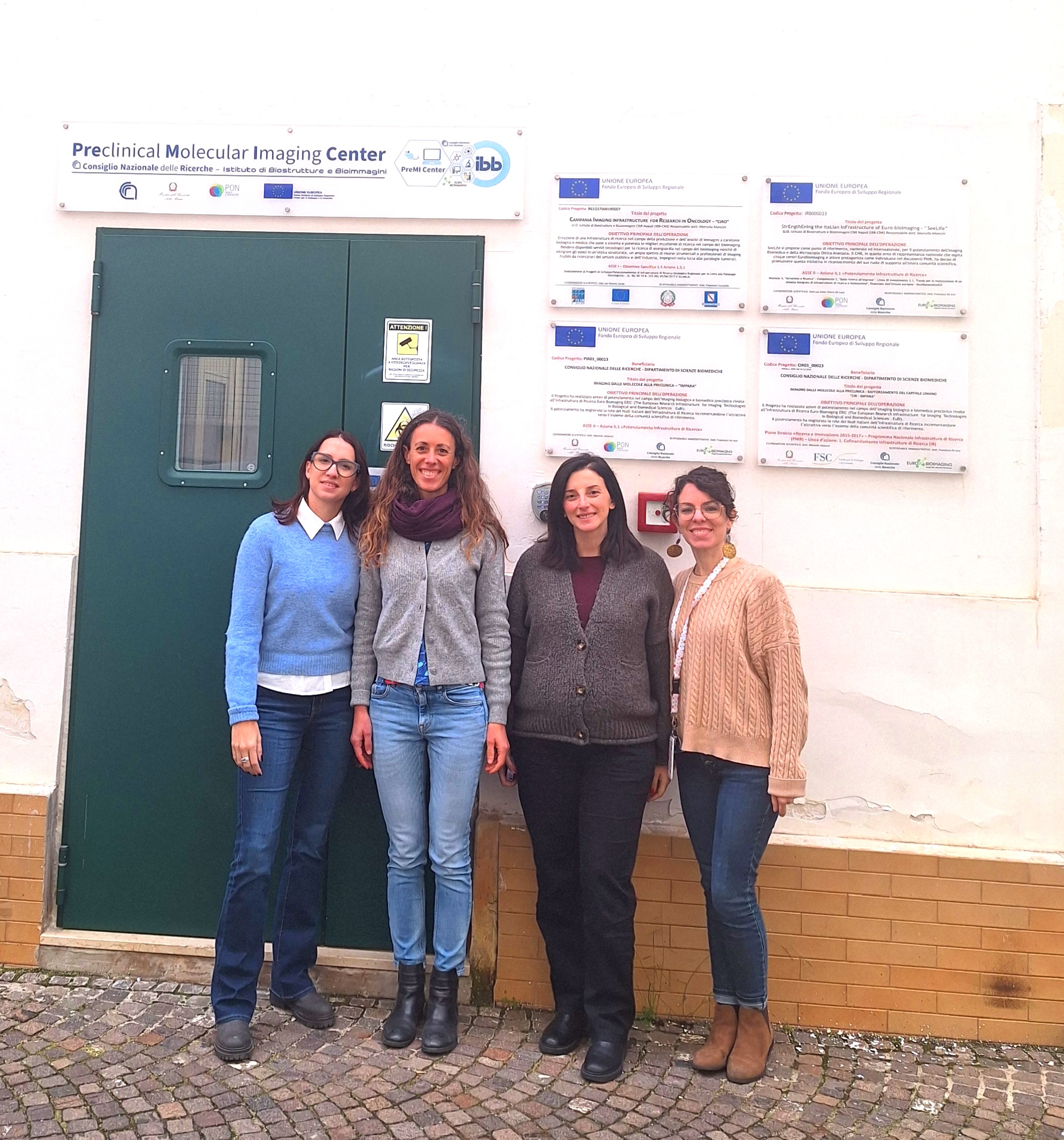
From left to right: Victoria Alonso and Erika Cerutti, members of the Euro-BioImaging Med Hub; Sandra Albanese and Serena Monti from the PreMI center during a visit to the node.
Supporting Researchers Across the Imaging Workflow
Both Serena and Sandra play key roles in helping researchers make the most of the facility's imaging capabilities.
Serena, a researcher at IBB-CNR since 2019, focuses primarily on magnetic resonance imaging (MRI) applications. Her role involves supporting and developing preclinical imaging studies, particularly MR-based applications, as well as assisting external users who access the infrastructure.
Sandra serves as Scientific Responsible for the PreMI Center. In her role, she coordinates imaging services and experimental workflows across the facility, promoting collaboration between experts in biomedical imaging, molecular biology, oncology, neuroscience, and cardiovascular research.
When researchers arrive at the facility, the team helps them select the most appropriate imaging modalities, optimize acquisition parameters, and develop data analysis strategies that ensure robust and reproducible results.
Expanding Capabilities Through New Technologies
In recent years, the PreMI Center has significantly expanded its technological infrastructure thanks to several major funding initiatives, including Italy's National Recovery and Resilience Plan (PNRR).
Among the new acquisitions, one technology stands out: a dissolution Dynamic Nuclear Polarization (dDNP) system. This cutting-edge technology enables dramatic enhancement of MRI signals from 13C-labelled probes, allowing researchers to visualize and quantify metabolic processes in real time.
Hyperpolarized MRI opens new possibilities for studying cancer, cardiovascular diseases, and other pathologies by providing functional and metabolic information that cannot be obtained with conventional imaging techniques.
We believe this technology has enormous translational potential and represents one of the most innovative tools currently available in preclinical imaging research.
Multimodal Imaging for Complex Biological Questions
The PreMI Center supports a wide variety of animal models, most commonly mouse models of cancer, cardiovascular disease, metabolic disorders, and neurological conditions. The facility also works with alternative models, including chicken embryos.
Serena’s expertise is primarily in magnetic resonance imaging, both preclinical and clinical, and image processing. She works with multiparametric MRI techniques, including anatomical, functional, diffusion, and metabolic imaging, as well as hyperpolarized MRI.
Sandra's expertise lies primarily in preclinical molecular and functional imaging, with a particular focus on High Frequency Ultrasound, PET/CT, Optical Imaging, and DEXA technologies. She shares with us her particular interest in tumor progression, angiogenesis, vulnerable atherosclerotic plaques, neuroinflammation, and brain functional activity. Sandra also contributes to ultrasound-guided procedures, micro-surgical interventions, and multimodal imaging studies aimed at enhancing the translational value and reproducibility of preclinical research.
This combination of expertise enables the facility to offer truly multimodal imaging approaches. Both researchers highlight the importance of integrating complementary information from different imaging techniques to obtain a more comprehensive understanding of disease mechanisms and treatment responses.
Sandra is particularly enthusiastic about high-frequency ultrasound:
This modality provides both morphological and functional information in a non-invasive and longitudinal manner, enabling us to characterize disease progression over time and evaluate the efficacy of novel therapeutic interventions.
Tackling Challenging Imaging Problems
One particularly memorable project involved developing an MRI workflow for imaging chicken embryos inside intact eggs.
Unlike conventional animal models, embryos can adopt different positions within the shell, making anatomical orientation difficult and preventing the use of standard reference planes. The team had to create flexible imaging protocols capable of identifying embryonic anatomy regardless of position or developmental stage.
The project required extensive methodological optimization but ultimately resulted in a robust and reproducible workflow for a model that is becoming increasingly important in developmental biology and translational research.
Building FAIR and Reproducible Imaging Research
The facility is actively working towards increasingly standardized and FAIR-compliant data management practices.
Imaging datasets are centrally stored within institutional infrastructures to ensure long-term accessibility and data integrity. Depending on project requirements and funding policies, data can also be shared through repositories such as XNAT to support reproducibility and data reuse.
Career Paths Shaped by Imaging
Although they arrived through different routes, both Serena and Sandra were drawn to imaging by its interdisciplinary nature.
Serena, a biomedical engineer by training, was attracted by the possibility of contributing to medical science through technological innovation and quantitative analysis. Her career gradually evolved towards MRI technologies, image processing, and translational imaging research.
Sandra began her career in a preclinical imaging facility at the University of Naples Federico II in 2012, gaining extensive experience in multimodal imaging and murine disease models before joining IBB-CNR. Today, she combines the scientific coordination of the PreMI center with the support of researchers by developing advanced preclinical imaging studies.
Looking Ahead
Both Serena and Sandra see the future of the PreMI Center as increasingly multidisciplinary and technology-driven.
Emerging fields such as hyperpolarized MRI, molecular imaging, artificial intelligence, and advanced image analysis are expected to play an increasingly important role in bridging basic research and clinical applications. At the same time, the facility aims to continue expanding its expertise and strengthening collaborations across scientific disciplines.
For young scientists considering a career in imaging, both have a similar message: stay curious, embrace interdisciplinarity, and gain hands-on experience.
My advice for young scientists would be to remain curious and embrace interdisciplinarity. Imaging is a field where biology, medicine, physics, engineering, and data science come together. Developing a solid scientific foundation is important, but being open to learning from different disciplines is equally valuable."
-- Serena Monti
I would also encourage gaining hands-on experience as early as possible, working in a multidisciplinary environment where physics, biology, and medicine intersect. This kind of exposure is what really helps you develop a translational mindset and understand how imaging can be applied to solve real scientific and clinical problems."
-- Sandra Albanese
-- Sandra Albanese
More news from Euro-BioImaging
July 15, 2026
Imaging-Driven Neuroscience, Responsible Research, and the Power of Advocacy: Highlights from FENS Forum 2026
Euro-BioImaging was pleased to participate in the FENS Forum 2026, Europe’s largest international gathering dedicated to advancing neuroscience (>8000 participants). Throughout…
Euro-BioImaging Highlights the Value of Research Infrastructures at the FENS 2026 Satellite Symposium hosted by EBRAINS
Euro-BioImaging was pleased to participate #FENS2026 Satellite Symposium “Accelerating Your Neuroscience Research through the European Research Infrastructures” organised by EBRAINS, to learn…