June 3, 2026
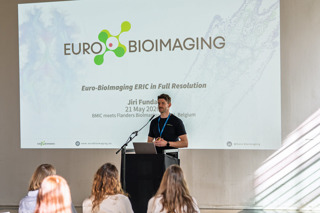
BMIC meets Flanders BioImaging
In May 2026, the Belgian Molecular Imaging Community (BMIC) and Flanders BioImaging joined forces in Ghent for a two-day community event that brought…

Euro-BioImaging is organizing an online User Forum on October 6, 2022, from 14:00-17:00 CEST. This event will highlight the importance of cutting-edge imaging technologies in support of fighting infectious diseases and showcase the specific expertise available at our Nodes across Europe through case studies presented in tandem with the research community.
At this event, learn how Cristina Risco Ortiz and her colleagues use live cell microscopy, correlative light and electron microscopy (CLEM), as well as three‐dimensional imaging methods to reveal how viruses manipulate cell organization to assemble their factories and shed new light on virus life cycle events (full abstract below).
Hear this talk and others like it on October 6 at the Euro-BioImaging User Forum: Fighting infectious diseases.

Imaging viral infections: Identifying new targets for antiviral treatments
Cristina Risco Ortiz, Cell Structure Laboratory, National Center for Biotechnology, CNB-CSIC, Madrid, Spain
As a result of recent advances in light and electron microscopy, we are starting to be aware of the variety of structures that viruses assemble inside cells. Viruses remodel cellular compartments to build replication factories. Remarkably, viruses are also able to fabricate new membranes and new organelles. Viral factories are intracellular compartments harboring viral replication complexes and the sites of assembly and maturation of new virus particles, that later move to organelles specialized in virus egress. Live cell microscopy, correlative light and electron microscopy (CLEM), and three‐dimensional imaging methods are unveiling how viruses manipulate cell organization to assemble their factories. In particular, methods for molecular mapping in situ in two and three dimensions, are showing how viruses build functional macromolecular complexes inside infected cells. The combination of all these imaging approaches is uncovering the viral lifecycle events with a detail never before seen and helping to identify new targets for antiviral therapies.
About Cristina Risco Ortiz:
Dr. Cristina Risco is the director of the Cell Structure Lab at the National Centre for Biotechnology in Madrid, Spain. Her group investigates the structural and molecular basis of virus-cell interactions and looks for new antivirals to combat Bunyaviruses and Coronaviruses.
To know more: www.cellstructurelab.es
References:
[1] Risco, C, Fernández de Castro, I, Sánz-Sánchez, L, Nayaran, K, Grandinetti, G, and Subramaniam, S. 3D imaging of viral infections. Annu. Rev. Virol. 1 (2014), 453-473.
[2] Fernández de Castro, I, Barajas, D, Fernández, JJ, Nagy, PD and Risco, C. Three-dimensional imaging of the intracellular assembly of a functional viral RNA replicase complex. J. Cell Sci. 130 (2017), 260-268.
[3] Fernández de Castro, I, Fournier, G, Sachse, M, Pizarro-Cerdá, J, Risco, C and Naffakh, N. A new class of Rab11-dependent vesicles transports influenza virus genome from a modified endoplasmic reticulum to the plasma membrane. Nat. Commun. 8, (2017), 1396.
[4] Tenorio R, Fernández de Castro I, Knowlton JJ, Zamora PF, Lee CH, Mainou BA, Dermody TS and Risco C. Reovirus σNS and μNS proteins remodel the endoplasmic reticulum to build replication neo-organelles. mBio 9 (2018), e01253-18.
[5] Sachse, M, Fernández de Castro, I, Tenorio, R, and Risco, C. The viral replication organelles within cells studied by electron microscopy. Adv. Virus Res. 105 (2019), 1-33.
[6] Fernández de Castro, I, Tenorio, R, Ortega-González, P, Knowlton, JJ, Zamora, PF, Lee, CH, Fernández, JJ, Dermody, TS, and Risco, C. A modified lysosomal organelle mediates the nonlytic egress of reovirus. J. Cell. Biol. 219 (2020), e201910131.
[7] García-Serradilla, M and Risco, C. Light and electron microscopy imaging unveils new aspects of the antiviral capacity of silver nanoparticles in bunyavirus-infected cells. Virus Res. 302 (2021), 198444.
June 3, 2026
In May 2026, the Belgian Molecular Imaging Community (BMIC) and Flanders BioImaging joined forces in Ghent for a two-day community event that brought…

June 1, 2026
The EOSC Association has adopted a new position paper calling for a distinct Work Programme-based Partnership for EOSC in FP10. Euro-BioImaging welcomes this…
May 29, 2026
Spatial transcriptomics is rapidly transforming the way researchers study complex biological systems. To explore the latest developments in this fast-moving field, Euro-BioImaging is…