Understanding how immune cells interact with different medications within the disease environment is at the heart of Alexandros Marios Sofias research interest. He is a principal investigator at RWTH Aachen University, whose lab focuses on understanding immune cells, designing nanomedicines, and targeting different aspects of the immune system. Multiscale imaging is crucial to his research because it enables him to assess what’s happening on the cellular, tissue and the full-body levels with different therapies. He recently visited Euro-BioImaging’s NORMOLIM Node in Trondheim, Norway, where he used very high temporal and spatial resolution intravital microscopy to visualize the tumor microenvironment in real-time. This visit was supported by the Euro-BioImaging pilot User Access fund.
Dr. Alexandros Sofias. Euro-BioImaging user & head of the “Immune Cell Targeting and Imaging” Research Group
A few years ago, Dr. Alexandros Marios Sofias and Dr. Sjoerd Hak identified the “hitch-hiking” concept in nanomedicine. This concept is based on the observation that specific targeted nanomedicines engage with specific myeloid immune cells in circulation (i.e., uptake of nanomedicines by circulating neutrophils and subsequent extravasation of the latter in the tumor microenvironment) thus providing a unique opportunity for drug delivery.
Observing the disease environment
“In the case of cancer, those immune cells are dragged into the disease environment,” explains Alexandros. “The fact that the drug is already inside the immune cells allows us researchers to “hitch hike” into the disease environment, and when observed on different scales, with imaging, we can better understand the delivery of nanomedicines to malignant / inflamed regions.”
In a complex project involving multiple research institutes, Alexandros and his team aim to specifically quantify the contribution of “hitchhiking” as a mechanism for delivering nanomedicine formulations to the tumor microenvironment in triple negative breast cancer.
The missing link - intravital microscopy
“In order to ensure that such a concept works on a clinical level, we need to perform multiscale imaging, on the cellular, tissue and whole-body levels,” explains Alexandros. “In Aachen we have excellent options for cell level and whole-body level imaging but we are missing the tissue level.”
In general, the ability to conduct intravital microscopy on mice is not commonly available in laboratories around Europe. But Alexandros knew that the Euro-BioImaging NORMOLIM Node in Trondheim, Norway, could provide it, since he had worked there during his PhD.
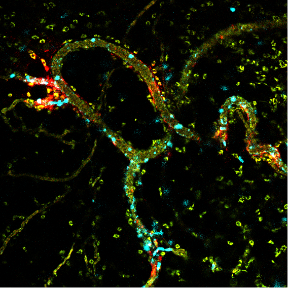
“High resolution intravital microscopy is the only available technique to enable real-time monitoring of nanoparticle-immune cell engagement. The Euro-BioImaging NORMOLIM Node in Trondheim offers an excellent intravital microscopy device at the moment - with fast detectors, strong lasers, and combination of confocal / multiphoton / second harmonic generation and expertise using it on mice,” explains Alexandros. “With this set up, we could visualize real-time nanoparticles and the immune cells and understand the interactions in circulation and in the tumor.”
Exemplary image of the tumor microenvironment visualized with intravital microscopy.
The Euro-BioImaging pilot User Access Fund provided the impetus for Alexandros to contact Dr. Sjoerd Hak, of the Norwegian University of Science and Technology, in Trondheim, Norway, with whom he had previously collaborated. The application was submitted in December 2021. He learned his application was successful in January 2022, and immediately started preparing for his trip.
“We would advise future users to establish contact with the Node beforehand; this will facilitate the entire process. Five to six months of scientific planning is necessary, especially for in vivo experiments,” he advises. “But finding an accommodation was relatively easy for me, I planned just a few months ahead of time. Sample preparation was also relatively straightforward. Some samples were prepared in Aachen, then sent to NORMOLIM with a courier company, while some samples were prepared in Trondheim.”
Alexandros’ first visit took place in June 2022. He stayed approximately one month, collecting a lot of data which he then analyzed at his home institute. He conducted a follow-up visit in January-February 2023. “The fact that the funding was flexible, allowing me to visit the facility twice, at a 6 month interval, was really efficient. I had a better focus the second time around,” he remarked. “And the team in Trondheim was very helpful, which is a definite time saver for developing in vivo models.”
Benefits of the project
This project was beneficial for Alexandros on many levels. “Scientifically, travelling to Trondheim to conduct these experiments was essential for raising the research quality,” says Alexandros. “And from a career perspective, I am really happy to have strengthened my skills and revived collaboration with Dr. Sjoerd Hak. I had worked with him in the past, but our collaboration had been dormant due to my move to Aachen. This was a nice opportunity to start collaborating again.”
“Euro-Bioimaging provides excellent opportunities for research exchange, facilitating the academic mobility for senior and young researchers. In addition, there are a lot of important actions taking place for informing scientists about the different possibilities,” concludes Alexandros.
Strengthening collaborations, honing researchers’ skills, raising the quality of research… The most important missions of Euro-BioImaging have been accomplished in this project. Thank you, Alexandros, for sharing your experience! We look forward to your publication!
More news from Euro-BioImaging
July 28, 2026
Global BioImaging Advance the Conversation on Sustainable Biomedical Imaging Facilities
How can biomedical imaging facilities remain scientifically excellent while becoming more financially, environmentally, and operationally sustainable? These questions were at the heart of…
Moara Lemos: New insights from Global BioImaging’s Job Shadowing programme
Moara Lemos is a Brazilian researcher and imaging scientist. Her award winning research pushes the boundaries of structural biology, using advanced cryo-electron microscopy…
A Multiplexed Vision: Highlights from the Special Edition Virtual Pub on Spatial Transcriptomics
On June 12th, Euro-BioImaging hosted its latest Special Edition Virtual Pub, bringing together over 200 researchers and technology developers working at the cutting…