Chronic intake of high amounts of fructose has been linked to the development of metabolic disorders caused by the almost complete clearance of fructose by the liver. However, direct measurement of hepatic fructose uptake is complicated by the fact that the portal vein is difficult to access.
Dr. Arjan Hendriks, from the University Medical Center Utrecht, applied for access at the PRIME Node to explore the opportunities and potential effectiveness of different deuterated compounds to map body metabolism with deuterium metabolic imaging (DMI), and thanks to the data obtained during this access he developed a new, non-invasive method to measure hepatic fructose uptake and metabolism, based on administration of [6,6’-2H2]fructose followed by DMI.
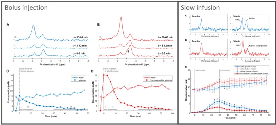
Tissue concentration of deuterated glucose and/or fructose, and of deuterated water as a function of time during and after a bolus injection versus a slow IV infusion of [6,6’-2H2]glucose and [6,6’-2H2]fructose. Figures have been adapted from: https://doi.org/10.3389/fphys.2023.1198578 Used under a Creative Commons CC-BY 4.0 license.
The application of this method in mice showed a higher extraction and faster turnover of fructose in the liver, as compared with glucose. Furthermore, comparing high concentration bolus injections with a slow low concentration infusion gave more insight into the fructose build up in the liver as compared to glucose. DMI with [6,6’-2H2]glucose and [6,6’-2H2]fructose could potentially contribute to a better understanding of healthy human liver metabolism and aberrations in metabolic diseases.
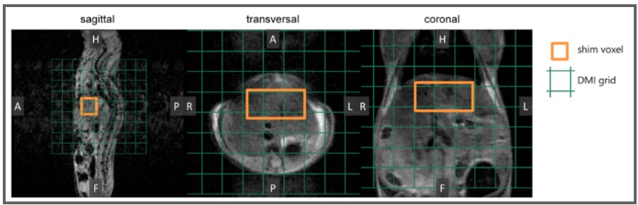
In vivo anatomical 3D MR images were acquired as a guide to perform B0 shimming on a voxel (orange) and to position the DMI grid (green). Figures have been adapted from: https://doi.org/10.3389/fphys.2023.1198578. Used under a Creative Commons CC-BY 4.0 license.
The Dutch Preclinical Imaging Center PRIME – Molecular Imaging Node is a dual-sited Node that consists of the Preclinical Imaging Centre (PRIME), Radboud University Nijmegen, and the Intravital Microscopy Facility (IMF), Hubrecht Institute, Utrecht. This Node is a centralized facility for multimodality imaging of small animals, such as mice and rats. PRIME is the only centre in the Netherlands where more than four imaging modalities for small animal imaging are housed under one roof in one facility. The intravital microscopy laboratory offers advanced genetic mouse models and has heavily invested in state-of-the-art imaging of small animals using multiphoton microscopy. PRIME functions as single-site facility for small animal imaging with state-of-the-art equipment and expertise for magnetic resonance imaging (MRI), positron emission tomography (PET/CT), single photon emission computed tomography (SPECT/CT), intravital multi-photon fluorescence imaging (MPFI) and high frequency ultrasound imaging (HFUS).
More news from Euro-BioImaging
July 23, 2026
Cellular Imaging Hungary Node expands expertise in advanced neurophotonics
The Euro-BioImaging Cellular Imaging Hungary Node has expanded its service portfolio with the addition of the BrainVisionCenter (BVC) in Budapest. Following a successful…
The UK Euro-BioImaging Node expands from seven to thirteen sites!
We’re delighted to announce that the UK Euro-BioImaging Node is expanding, growing from seven to thirteen sites following a successful upgrade application and…
Building Skills Across Europe: EVOLVE Supports the Second Edition of the Distributed Image Analysis Training Course
The second edition of the “Introduction to Image Analysis with Python for Life Scientists” course marked another successful milestone in the development of Euro-BioImaging’s distributed training model.