Looking at Drosophila development with laser microdissection & 2-photon microscopy
Published November 21, 2023

Country
Node
Technology
Category
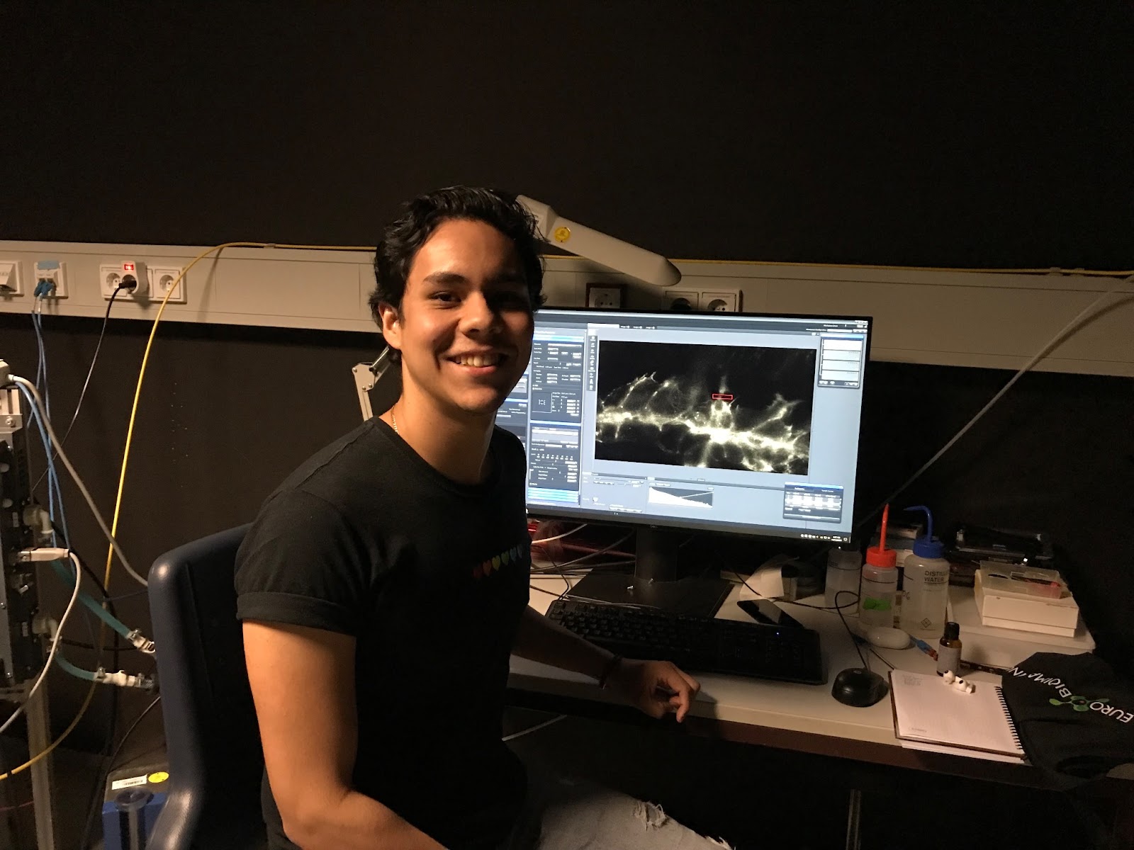
Daniel Ríos Barrera is a developmental biologist and Associate Investigator at the Instituto de Investigaciones Biomédicas (IIBO) of the Universidad Nacional Autónoma de México (UNAM). In his lab, Daniel and his team study how tubular systems are formed, focusing on the development of the respiratory system in fly embryos as a model for more complex organisms like humans. His quest to understand these developing organs brought him and his PhD student, Luis Eduardo Sánchez Cisneros, to EMBL’s Advanced Light Microscopy Facility, part of Euro-BioImaging’s EMBL Node. At EMBL, they will use laser microdissection and 2-photon microscopy to observe the recoil of the developing tissues. A quick discussion with Daniel & Luis reveals how access to EMBL’s technologies and expertise will help them answer their scientific question, and how it may impact their careers.
Fruit fly embryos are a common model system for developmental biology. “With fly embryos, most of the branching of tubular tissue can be observed in just 2 hours,” explains Daniel. “These tissues are particularly interesting because they bring oxygen directly to tissues, instead of using blood to carry out this function. In our home lab, we do live cell imaging with confocal live microscopy, looking at fluorescent proteins and parts of the cells. But we would like to have a better understanding of the mechanical properties of the entire tissue, not just on a cellular level.”
Understanding the development of tubular systems
“That’s why I applied to visit Euro-BioImaging’s EMBL Node. EMBL has microscopes that will allow us to manipulate the tissue using laser microdissection. In addition, EMBL staff are experts in the biomechanics field. They will be able to help us understand the force and recoil of the tubular systems we are interested in. Furthermore, the ALMF facility offers s several alternative instruments to perform these experiments, such as two-photon microscopy where we can cut tissue using infrared light, or a system which uses UV light but is more versatile than the two-photon.”

“We’ve already tried both systems. And the 2-photon system is very promising,” explains Daniel. “Luis has been trained on these systems now. As these machines require a very different approach to the ones we have at home, we’re glad he has picked up this new skill.”
Learning from the experts
While Daniel did his postdoc at EMBL, this is Luis’s first time visiting, and Daniel is excited for his student to have this experience and exposure.
“This is my first experience abroad,” says Luis, who is in the first year of his PhD. “I was very excited, but also very nervous, before I left home,” he said shyly. “I was nervous about my English. I also thought EMBL would be very intimidating. It is so big and there is so much going on,” he explained. But all of his fears were quickly dispelled when he arrived. “There is such a friendly atmosphere here. People are collaborating across groups - it’s really nice.”
Even the microscopes were less complicated than he thought they would be. “The microscope systems I am using here are intuitive and user-friendly. And with these microscopes we can do much more than we can in our home lab.”
The trip to EMBL lined up nicely with the European Drosophila Research Conference, a major event for Drosophila researchers taking place in Lyon, France. “Lyon is just a short train ride from Heidelberg, when you’re from Mexico,” jokes Daniel. “Travelling to Europe from Mexico is very expensive,” explains Daniel. “Grouping this conference with our research work makes it possible to do both. It’s wonderful that this has worked out.”
For Daniel and Luis, the conference was a wonderful opportunity to meet with their peers, exchange ideas, show their results, and get feedback. It was Luis’s first time to attend a Drosophila-specific conference and his poster was very well received. “There were 100s of posters, but still, many people stopped by to talk to us,” he says with a smile.
Daniel returned to Mexico after the conference, but Luis will stay at EMBL for several more weeks, continuing to generate images. In the end, it works out nicely, because he will be able to participate in the EMBL PhD Symposium before he returns home.
“At first, I hesitated, because of my English. But I can’t let this opportunity pass me by. I will present our project poster,” he says proudly.
“It’s really exciting to be able to undertake this project,” says Daniel. “Being a Euro-BioImaging user allows me to stay in contact with the people I worked with previously at EMBL, while sharing new skills with my students.”
And Luis has enjoyed learning from the experts at the ALMF. “I’ve learned how to use the microscopes but I’ve also learned the theory behind them. In addition, I now know how to do the data analysis, and image processing with Fiji, skills I will take back to Mexico. And finally, I learned that things aren’t as intimidating as I expected. From the application process at Euro-BioImaging, to working with the experts at EMBL, to presenting my work in English, everything has been easier than I thought.”
As the project advances, they will work with a partner in France, Philippe Bun, from the NeurImag Imaging Facility, IPNP (Inserm U1266, Paris), part of the French Node of Euro-BioImaging, to delve further into the image analysis. So the project continues, with an international flair.
Daniel commented that he is also glad that the Euro-BioImaging spirit has now spread to other parts of the world including Mexico. In Mexico, the Mexican BioImaging and Latin America BioImaging networks, dedicated to training imaging scientists, are very active. Daniel recently participated in one of the workshops that Mexican BioImaging organized in the institute where Daniel did his PhD, and next year, Daniel’s current institute will host another course of the Mexican Bioimaging network. In September 2023, Mexico BioImaging became an official community at the joint Latin American BioImaging/BioImaging North America meeting and is now part of the larger Global BioImaging community.
More news from Euro-BioImaging