How does prostate cancer become aggressive and life-threating? That is a question that Maria K. Andersen, post-doc researcher at the Norwegian Institute of Science & Technology (NTNU) - Trondheim, really wants to answer. She is part of the ERC-funded ProstOmics project, which combines -omics technology with state-of-the-art imaging approaches to better understand the molecular mechanisms underlying prostate cancer development and validate biomarkers and molecular signatures for separating aggressive from indolent prostate cancer. She believes cholesterol plays a role - but it is very difficult to detect cholesterol in tissue using the conventional mass spectrometry techniques she works with in her lab. To further explore the role of cholesterol in prostate cancer, she applied for access to Euro-BioImaging’s Facility of Multi Modal Imaging AMMI Maastricht Node, where a highly sensitive MALDI2 MSI instrument is available in open access. Maria tells us about her experience.
“There is already a lot of research in the role of cholesterol in prostate cancer, but most of it is related to serum levels. There are very few studies looking at cholesterol detection in tissue,” explains Maria. “But I think it’s really important to map what actually happens with cholesterol in prostate tissue. Unfortunately, it’s not possible to do so with MALDI Mass Spectrometry Imaging (MALDI MSI), the technology we use in the ProstOmics project. But MALDI 2 allows you to detect sterols (like cholesterol). The idea behind my pilot project is really to develop and validate an approach using MALDI 2 that can be used on a wider-scale to help understand the molecular mechanisms underlying prostate cancer development.”
To do so, she needed the additional expertise of the M4I group in the Netherlands, part of our Facility of Multi Modal Imaging AMMI Maastricht Node. She had heard about the Euro-BioImaging pilot User Access fund from a colleague and decided to apply, since the ERC funding does not cover this type of additional experiment.
She learned her application was successful in February 2022 and immediately made arrangements for her visit to the AMMI Maastricht Node, a facility where she had already conducted some experiments in the past.
Prior planning for experiment success
“I suspected the instrument would be in high demand, so I made my request 4 months ahead of time. I also had to find accomodation which would allow me to stay near the facility for 9 consecutive days. It was really important for me to be well ahead of time and be able to communicate with the core facility staff about the instrument bookings.”
Upon arrival in Maastricht, she tested the different matrices, to see what worked best, in collaboration with the Node staff, who gave some advice and ideas on what to test. Then she did the actual imaging work - she was able to image 32 samples in merely 2 days. Then she assessed and analysed the images with the help of the experts at M4I.
“I was in the Netherlands for 9 very intense days,” says Maria.
Outcomes
“I’m really happy with the results. I was able to detect cholesterol and a few other sterols. I hoped to be able to detect intermediates – but that wasn’t feasible as the levels were probably too low in the tissue. We will definitely publish our results soon and further work on mapping these results to the other multi-omics analysis we have, including proteomics, transcriptomics, metabolomics, lipidomics and epigenomics.”
“The assistance I got from the M4I Node personnel with planning, execution and data interpretation was crucial for the success of this project,” explains Maria. In particular PhD-fellow Britt Claes and researcher Michiel Vandenbosch provided excellent expertise and help both before and during her stay.
“So I would like to return to the Node, to try this approach on 200 more samples. I would need a bigger sample size to really be sure of the biological meaning of the results – especially if I want them to translate to the clinic. So I’ve applied for a new grant, this time to get enough funding for a 6 months research stay and instrument time.”
Future perspectives
Maria is at the end of her post-doc, and she would like to continue working as a researcher. Getting the funding from the Pilot User Access fund has been helpful in building her CV. Thanks to this fund, she has not only learned a new technique, but also gained a stamp of approval for her work, especially with regards to further funding applications. And most importantly, this project has contributed to building a long-lasting collaboration with her colleagues in Maastricht.
And that after all is one of the important missions of Euro-BioImaging, to build the scientific collaborations that lead to excellent science in the European Research Area.
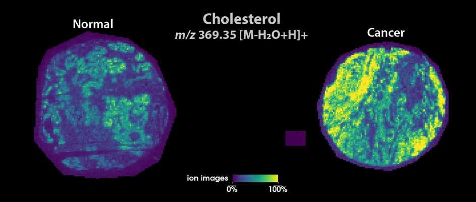
Spatial distribution of cholesterol on a prostatic normal (left) and a cancer (right) sample.To spatially detect sterols, Maria used a timsTOF Flex instrument coupled with a MALDI-2 source at the Facility of Multi Modal Imaging AMMI Maastricht Node.
More news from Euro-BioImaging
July 28, 2026
Global BioImaging Advance the Conversation on Sustainable Biomedical Imaging Facilities
How can biomedical imaging facilities remain scientifically excellent while becoming more financially, environmentally, and operationally sustainable? These questions were at the heart of…
Moara Lemos: New insights from Global BioImaging’s Job Shadowing programme
Moara Lemos is a Brazilian researcher and imaging scientist. Her award winning research pushes the boundaries of structural biology, using advanced cryo-electron microscopy…
A Multiplexed Vision: Highlights from the Special Edition Virtual Pub on Spatial Transcriptomics
On June 12th, Euro-BioImaging hosted its latest Special Edition Virtual Pub, bringing together over 200 researchers and technology developers working at the cutting…