In this edition of our Meet the Nodes interview series, we introduce Irma Mahmutovic Persson, staff researcher at the Lund BioImaging Centre (LBIC), Lund University, part of the Euro-BioImaging Node Swedish National Microscopy Infrastructure (NMI). Working across both the preclinical MRI and nuclear medicine platforms, Irma plays a central role in coordinating in vivo imaging studies and supporting researchers throughout the experimental workflow.
A versatile role across imaging platforms
Irma joined LBIC in 2021 and splits her time between the 9.4T preclinical MRI platform and the nuclear medicine platform, working with SPECT-PET-CT scanners. Her role varies significantly depending on the project, reflecting the multidisciplinary nature of core facility work.
She contributes to study design, logistical planning, animal handling, and data acquisition. In some projects, she supports researchers with specific technical procedures such as radiotracer injections, while in others she is involved from hypothesis development to final data analysis.
She mainly contributes through study planning and project management, helping ensure that experiments are designed with proper controls, standardized timing, and optimized imaging strategies.
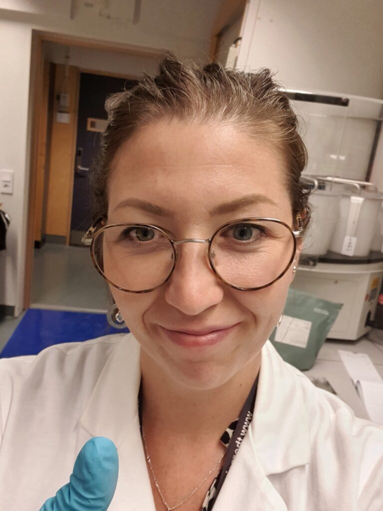
Irma Mahmutovic Persson at LBIC
Supporting a diversity of research questions
Irma primarily works with preclinical MRI and PET-CT, while also contributing to SPECT studies when needed. Her work involves advising researchers on protocol optimization, tracer timing, and experimental reproducibility, all key aspects when studying disease progression over time.
LBIC has supported and continues to support a wide range of disease models using diverse imaging approaches, including:
Stroke and traumatic brain injury, studied using advanced MRI techniques such as diffusion imaging and tractography
Glymphatic system and brain connectivity studies, primarily investigated through high-field MRI
Tumour theragnostics using radionuclide tracers, monitored using SPECT-CT and PET-CT imaging
Lung fibrosis, inflammation, and ventilator-induced lung injury models, assessed mainly with PET-CT and complementary CT imaging
Bone regeneration and skeletal imaging, supported through PET imaging and high-resolution micro-CT
Neonatal brain haemorrhage models, longitudinally monitored using SPECT-CT and MRI workflows
A recent example of advanced multimodal research at LBIC involves glioblastoma models, combining MRI-guided focused ultrasound treatment, contrast-enhanced MRI, and SPECT imaging within the same animal to monitor therapy response.
Favourite technologies and challenging workflows
Irma particularly enjoys working with MRI and PET imaging. She highlights MRI’s ability to provide detailed anatomical and structural information, combined with PET’s strength in monitoring functional and molecular processes over time.
One of the most technically demanding projects she recalls involved scanning animals in vivo using MRI, followed by high-resolution ex vivo MRI of extracted organs and subsequent light-sheet microscopy imaging, at the LBIC microscopy platform. Coordinating workflows between platforms, maintaining strict timelines, and ensuring sample integrity required extensive cross-facility collaboration.
There was no space or time for mistakes! And of course, we had some air bubble issue appearing in the liquid for some of the organ scans ex vivo, which we needed to deal with. Fun challenge, and personal development at most, even though it was challenging for us to perform it, I enjoyed the encounter.
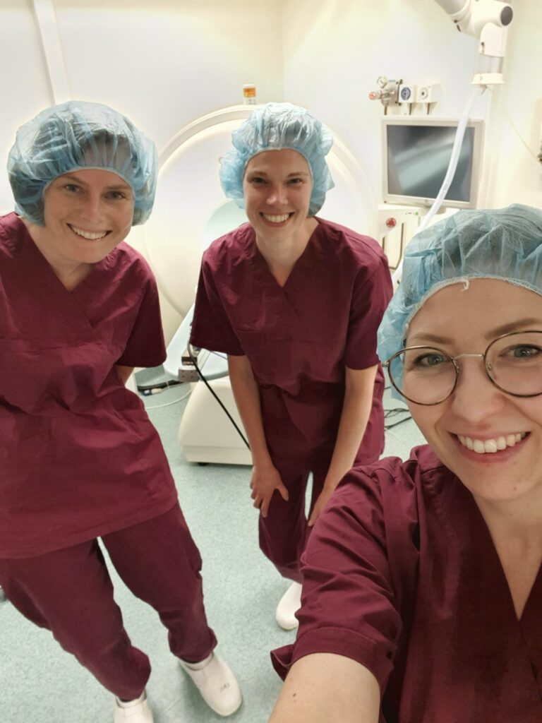
Irma and collaborators from Radboud University Medical Centre, Nijmegenduring a visit to LBIC.
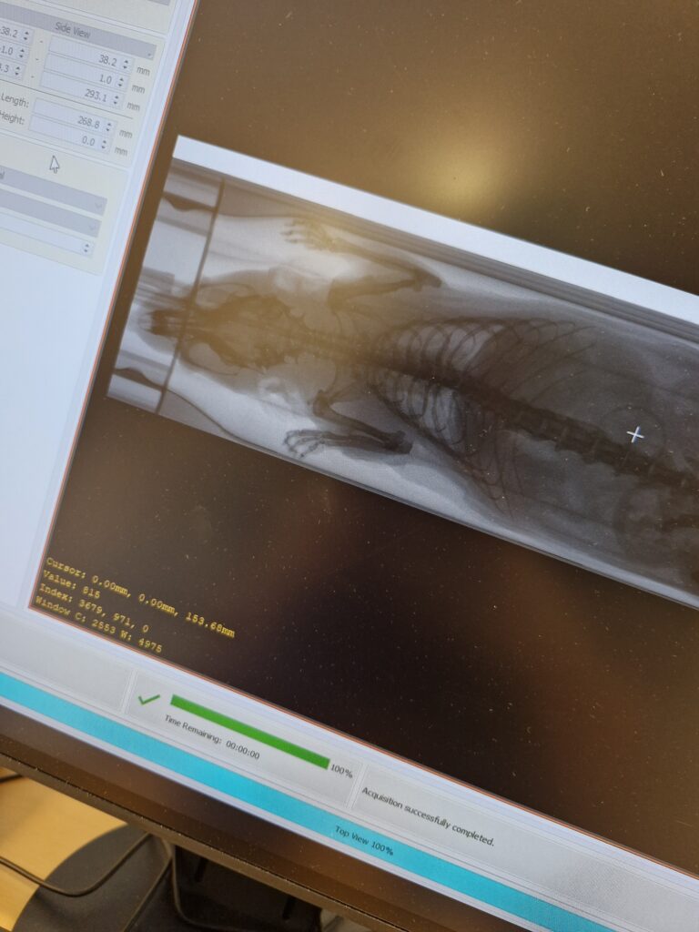
Rat CT scan part of a project investigating Ventilation-induced lung injury to study tissue strain in vivo.
From biology to multimodal imaging
Irma’s academic background began in molecular biology, followed by a PhD in respiratory immunopharmacology at Lund University, where she studied lung inflammation across multiple biological scales.
She later expanded into imaging through her postdoctoral work in the European TRISTAN consortium, focusing on translational imaging for drug safety assessment. Working extensively with PET-CT-MRI multimodal imaging during this period strengthened her technical expertise and collaborative network across Europe.
Her experience as a frequent user of LBIC during her postdoc ultimately led to her current role at the facility.
Future directions for LBIC
LBIC is currently undergoing structural transformation, merging with other infrastructures within Lund University to form a larger integrated research unit. This development is expected to expand collaboration opportunities and increase the impact of imaging across diverse scientific disciplines.
Recent upgrades across LBIC platforms, including microscopy, clinical scanners, and preclinical imaging systems, are already enabling: higher image resolution, faster acquisition times and more precise and reproducible workflows. Irma notes that interest in bioimaging continues to grow among researchers, reinforcing the importance of advanced imaging infrastructures.
I have noticed increasing interest of bioimaging in general among researchers, which is a nice trend to be part of!
Looking ahead: personal and technological growth
Irma’s responsibilities have expanded since joining LBIC. She now manages in vivo experimental workflows, ethical approvals, animal logistics, and facility administration related to animal research. Future growth and development might entail additional expertise in operating the modalities with new applications and novel sequences or tracer that haven’t yet been assessed at the facility yet.
I wish to learn more about the physics behind the scanners and detectors in order to be able to push the technologies further, in the search for better resolution or development of early biomarkers within disease progression models.
Advice for future imaging specialists
Irma encourages early-career scientists and technicians to pursue imaging regardless of their academic background:
Just go for it! It does not matter if you come from the biology field and aim to learn the technologies or the physics, or the other way around. If you think it is possible, make sure to try and do not give up the first time someone tells you it is impossible.
She recalls being told that MRI and PET would not be possible to run consecutively on two different scanners, within the same animal, and merge the images to answer my research question. But she made sure to increase her odds, and developed phantoms, 3D-printed modules to enable docking of different animal beds onto another scanner from a completely other vendor, and figured out the geometrical association to overlay the images, leading to proud published work within multi-modality imaging.
Finally, a reflexion on what she enjoys the most about her job:
I get to be part of exciting journeys and stories in each and every project I get involved in, and the best part is that it never gets boring. No day is similar to the other! Not to mention the exciting results I get to be part of and answer fascinating research questions.
More news from Euro-BioImaging
April 9, 2026
Major EU funding for user access & AI development, staff training, data stewardship & many more exciting new services!
Euro-BioImaging ERIC is deeply grateful to announce that the European Union has entrusted our infrastructure with funding to shape the future of imaging and…
Euro-BioImaging is looking for an Operations Support Assistant at the Euro-BioImaging Statutory Seat in Turku, Finland, to support the day-to-day financial and administrative operations…