April 13, 2026
We are hiring a Data Architect/Knowledge Engineer
Euro-BioImaging Bio-Hub team, hosted by EMBL in Heidelberg, is hiring a Data Architect/Knowledge Engineer – Euro-BioImaging (AI4Access) to…

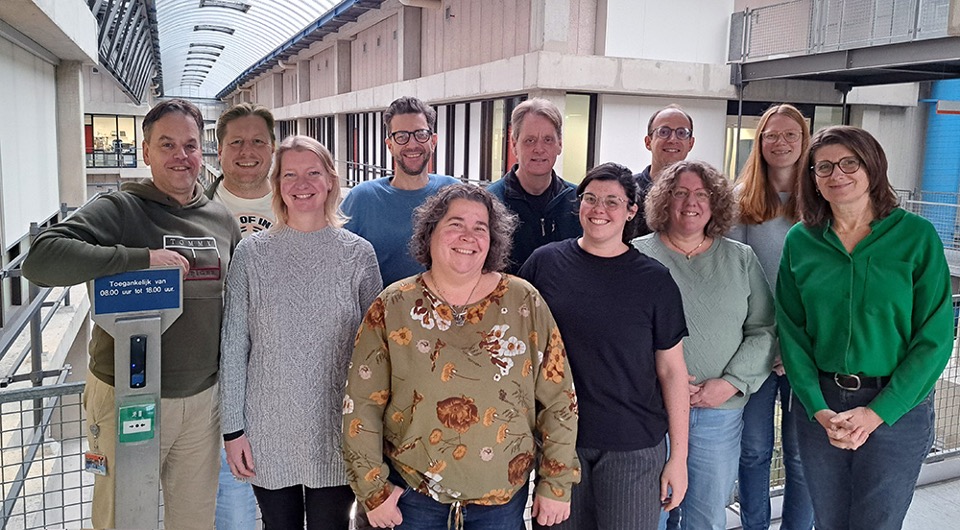
As part of our ongoing series spotlighting the scientists working at imaging facilities across the Euro-BioImaging realm, we sat down with Przemek Krawczyk, a microscopy specialist and group leader at the ‘Advanced Light Microscopy, Flow Cytometry and Electron Microscopy Center Amsterdam’ at the Amsterdam UMC, part of NL-BioImaging. Przemek brings a combination of scientific knowledge and technical expertise to the facility. Today, he guides users across a broad spectrum of imaging techniques, from high-content automated microscopy to live-imaging. At the same time he is leading the development of BIOMERO, an open-source platform to reshape how researchers store, share, and specifically, analyze imaging data.
By Lieke Stockmann, Euro-BioImaging Scientific Ambassador, University of Copenhagen
Przemek's intersection with the core facility was not a sudden shift. In parallel with running his own research group, he gradually took on a more formalized role within the core facility, a move that allows him to bring his biological and technical expertise to a wide imaging user community.
The core facility where Przemek works operates across three main pillars: electron microscopy, light microscopy, and flow cytometry. He is embedded in the light microscopy section, where he supports users with a range of techniques while drawing on his hands-on experience as a research biologist to help troubleshoot experiments and design workflows.
Beyond the facility itself, Przemek is involved in teaching and training the wider imaging community. Together with a partner institute in Amsterdam (NKI), the facility runs both introductory and advanced microscopy courses for graduate students.
Przemek has a pragmatic view to imaging and the choice of imaging instruments. "I see imaging techniques as tools, and the choice of tool depends on the question you want to ask," he says.
"Research is moving more and more towards automated microscopy and big data. The different omics techniques are becoming more popular, they're reaching single-cell resolution, and multi-omics approaches are being developed left and right."
-- Przemek Krawczyk, Microscopy specialist and Group leader, Amsterdam UMC
Therefore, the facility's portfolio of instruments spans the spectrum: from automated platforms such as the Satorius Incucyte and Molecular Devices ImageXpress Pico, to semi-automated systems like the Thunder microscope. The latest addition to this spectrum are the Leica Cell DIVE, a multiplex imaging system, and a laser microdissection microscope. Przemek is involved with these systems in different capacities from advising users to building computational pipelines that tie them together.
His technical ambition is not limited to commercially available equipment. Earlier in his career, he built his own custom confocal microscope coupled to an ultrasoft X-ray source, which he mentions as one of his greatest technical challenges he worked on.
A very exciting avenue Przemek is focusing on today is connecting multiple technologies into a single workflow that can extract richer biological information from complex samples.
At the center lies the Leica Cell DIVE system, an immunofluorescence microscope capable of imaging the same sample with up to 60 different antibodies. The concept is analogous to spatial transcriptomics, but using antibodies instead of probes. "It's actually a relatively simple microscope with only four channels," Przemek explains, "but the idea is that you label your cells using a robot, strip the antibodies, and repeat the process.”
The ultimate goal is to link this rich spatial protein data to downstream molecular analyses. Once regions of interest are identified from the multi-marker images, they can be dissected using the laser microdissection microscope and used for omics analyses. Here you can think of transcriptomics, lipidomics, metabolomics and with the advances these days this could lead to information at the level of individual tissue regions or even single cells.
The challenge is how to string it all into a single workflow. "Every part is challenging because it turns out it's not so easy to label your cells with antibodies sequentially,” Przemek says, “And then to flawlessly connect it to the micro-dissection and to the omics downstream.” The team is currently running pilots and establishing pipelines and protocols towards this single workflow.
More broadly, the project reflects where biology and the imaging community is heading. "Research is moving more and more towards automated microscopy and big data," he notes. "The different omics techniques are becoming more popular, they're reaching single-cell resolution, and multi-omics approaches are being developed left and right. This project fits within that bigger picture."
Alongside his work at the core facility, Przemek co-leads development of BIOMERO. BIOMERO is an open-source toolkit that augments the popular image storage platform OMERO. A second paper describing the latest developments is already available on ArXiv.
"OMERO is very limited in its current form," he explains. "We're trying to convert it into a future-ready tool." The goal for BIOMERO is to handle the complete lifecycle of imaging data: storing images, sharing them, running analyses, training machine learning models, and ultimately enabling GenAI-powered pipelines that can guide researchers through complex workflows.
The platform is funded through a grant from the Dutch Research Council (NWO), and Przemek works with a team of engineers to build it. They started rolling it out internally at the Amsterdam UMC and then across other institutions in the Netherlands. A national instance serving all Dutch researchers is expected to launch soon, with ambitions to attract international users as well. What makes BIOMERO particularly attractive is its approach to AI integration. The team has already established the technical foundations for interactions between AI and OMERO. "Think of uploading an image or a dataset, then training a machine learning model for segmentation on your own data," Przemek describes. "Or having an AI assistant help you figure out a more complex processing pipeline for a single image, converting that pipeline to code and running it on a compute cluster with GPU resources to analyze much larger datasets. Then getting results back and using the assistant to help extract the information you actually need from the data tables."
To conclude, we asked for some career advice for those interested in following a similar path at the intersection of microscopy and AI. This field is less about a clearly defined trajectory and more about navigating a rapidly evolving hardware and software landscape. Przemek answer is therefore clear: “If you want to keep up with AI, use AI. No one has any any real advice at the moment, since there are no true experts. The best way to become one is to do it.”
At the same time, a solid scientific background remains important. For now, a bachelor’s or master’s degree, ideally combined with internships or hands-on exposure, is still the best entry point. However, motivation and curiosity can take you a long way. In environments such as core facilities, enthusiasm and a willingness to learn often matter as much as prior experience.
April 13, 2026
Euro-BioImaging Bio-Hub team, hosted by EMBL in Heidelberg, is hiring a Data Architect/Knowledge Engineer – Euro-BioImaging (AI4Access) to…

April 9, 2026
Euro-BioImaging ERIC is deeply grateful to announce that the European Union has entrusted our infrastructure with funding to shape the future of imaging and…
April 8, 2026
Euro-BioImaging is looking for an Operations Support Assistant at the Euro-BioImaging Statutory Seat in Turku, Finland, to support the day-to-day financial and administrative operations…