
July 9, 2026
Euro-BioImaging Visits the NORMOLIM Node at the 2026 180°N Conference
In April 2026, the Med-Hub Head of Operations Alessandra Viale attended the 2026 180°N Conference, held at the beautiful Nye Hjorten Teater…

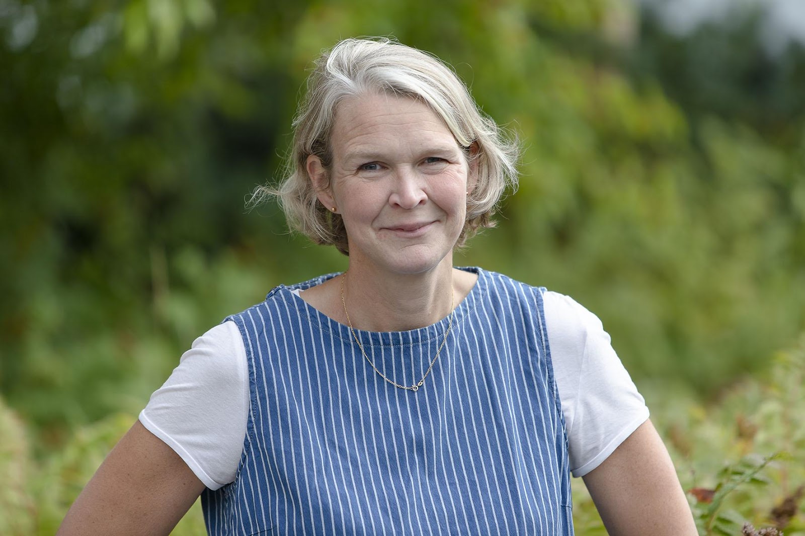
Delivering cutting-edge Electron Microscopy (EM) support, operations, and instrumentation, that is the mission of the Umeå Centre for Electron Microscopy at Umeå University, part of Euro-BioImaging’s Swedish NMI Node. It’s a relatively new facility, with a range of imaging techniques, from CLEM to cryo-EM. In order to support rapid developments in the cryo-EM and Electron Microscopy fields, and better serve a widening user base with diverse applications in structural and cell biology, plant science and infection medicine, Linda Sandblad, the Director of the Umeå Centre for Electron Microscopy, saw a growing need for method development, to establish the project workflows & data analysis pipelines that will ensure state-of-the-art service provision in the future. So, she applied for the prestigious Research Infrastructure Fellow grant from the Swedish Foundation for Strategic Research (SSF) – and got it. We spoke to her to see how this grant will support the establishment of pioneering methods to get the best results out of the novel technologies available at her facility.
“As the Director of the Umeå Center for Electron Microscopy, I lead the organization and its development. Currently, we are a staff of 9 people working with electron microscopy or correlative microscopy. We support researchers from Umea and beyond,” says Linda Sandblad. “We have developed a complete, one-of-a-kind suite of instrumentation for advanced EM projects that is unique in Sweden. This provides an excellent basis for method development and/or to initiate creative interdisciplinary research projects.”
The facility is part of the SciLifeLab and Swedish NMI, the Euro-BioImaging Node. Through Linda’s research activities at Umea Center for Microbial Research/Molecular Infection Medicine Sweden, it is also part of the EMBL Nordic Partnership. Within the EMBL Nordic partnership, the facility supports correlative microscopy projects, providing image analysis, mainly for cell, microbiology and infection biology.
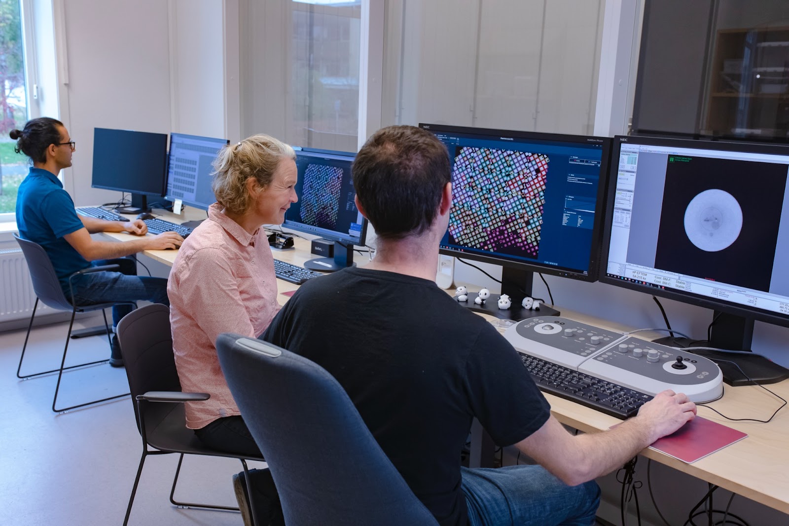
“We are fortunate to have state-of-the-art instruments at our facility,” says Linda. “In 2022, we purchased a Cryo-EM instrument with a new and really fast electron detector: Falcon 4i and micro-electron diffraction for cryo-EM data collection of small crystals. We currently have seven electron microscopes. Also, our Titan Krios Cryo-EM microscope were just upgraded with a Selectris-Falcon4i energy filtered detector. We also have top-of-the-line sample preparation instruments to do correlative projects, combining light microscopy with electron microscopy and tomography. This allows us to work on cell biology and infection biology applications.”
“Now we need to perfect our methods and develop our image analysis expertise. This is crucial for the for interdisciplinary & correlative imaging projects we will support in the future,” says Linda.
With 1,5 Million Euro over the next five years, Linda Sandblad will invest more in human resources for method development. In addition, the funding she received can be used for instrument service and consumables.
Sample preparation
Another area she will invest in is sample preparation. Currently, the facility sample prepration labs are open for local facility users and visitors, as a collaborative space. Now, they build on bacteria and cell culture facilities, to be able to work close to both live cell imaging and different cryo-fixation methods, such as high pressure freezing for tomography and cryo-EM. “It is tricky, but we think we can win more in following the cells we can see with fluorescence, when we know they are happy before we cryo-fix them, to follow the correlation all the way to structural level by electron microscopy” explains Linda.
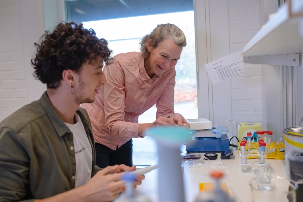
Image analysis pipelines
The research community has moved into the area of Data Driven Life Science (DDLS). EM volume imaging of cells and tissue, display the full content of the entire biological system present in the specimen after sample preparation. As EM image quality and resolution improves, new applications are possible, including proteomics, metabolomics and lipidomics methods, displaying cellular content with other types of data. DDLS will provide the capability of merging data from different research disciplines and creating new possibilities.
“There are a lot of very nice academic developments going on all over the world. We’re not going to build new software but rather concentrate on making existing tools more accessible to the inexperienced. Today, it is very tricky for beginners to get started with existing image analysis tools. We want to simplify use of the good tools that exist and develop the visualization of correlation of microscopy and interdisciplinary data,” explains Linda.
New instruments, new methods
“I hope that we manage to find really effective workflows. The challenge is that every biological model is different, every cell is unique, every project needs optimization and experience. We want to find good methods we can advise to researchers.”
“Of course,” adds Linda, “These investments will benefit all scientists in Europe. Visitors come from all over for more long-term visits, weeks or months to use our facility. We can get so much more out of our facility resourses when we have time to optimize the methodology for a specific project.”
About Swedish NMI
The National Microscopy Infrastructure (NMI) is a distributed infrastructure of five specialized, complementary, and interlinked sites located across Sweden: Stockholm, Uppsala, Göteborg, and Umeå. The mission of NMI is to provide access to innovative technologies and competence in microscopy and image analysis to all scientists that have a need for advanced imaging in fundamental and translational biomedical research projects. Annually, the NMI serves more than 500 national and international users. The Swedish Euro-BioImaging Node has strong expertise in super-resolution microscopy, correlative and multimodal microscopy, intravital microscopy as well as image processing and analysis. The infrastructure is coordinated by KTH (Royal Institute of Technology) which provides the single-entry point from which users are directed to the relevant imaging technologies.
July 9, 2026
In April 2026, the Med-Hub Head of Operations Alessandra Viale attended the 2026 180°N Conference, held at the beautiful Nye Hjorten Teater…
July 7, 2026
Armed conflicts generate long-lasting environmental contamination that extends well beyond the duration of military operations. The release of heavy metals such as Arsenic,…
July 6, 2026
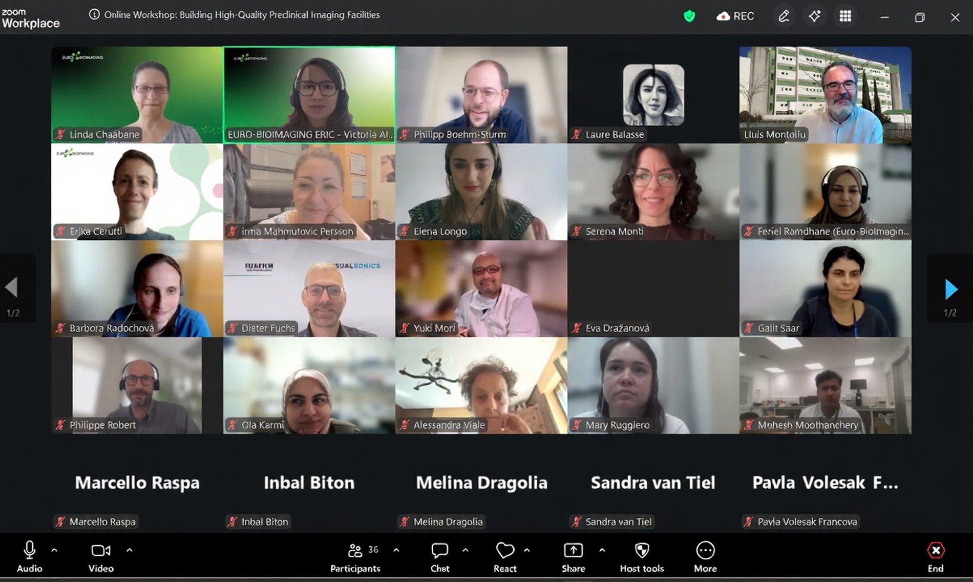
On 2 July 2026, Euro-BioImaging hosted the online EVOLVE workshop “Building High-Quality Preclinical Imaging Facilities”, bringing together approximately 40 imaging facility staff and…