MINFLUX is a super-resolution approach developed by Nobel prize laureate Stefan Hell in 2016. Two Euro-BioImaging Nodes are currently offering MINFLUX in open access as part of our Proof-of-Concept study. We spoke to Timo Zimmerman, Head of Light Microscopy applications at the EMBL Imaging Centre, part of Euro-BioImaging’s EMBL Node, to learn more about this technology and what it can be used for.
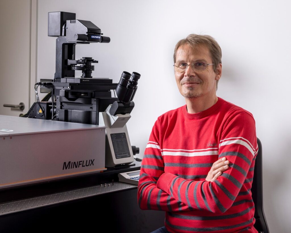
Timo Zimmerman, team leader for light microscopy services at the EMBL Imaging Center. Photo courtesy of EMBL PhotoLab.
MINFLUX is the super resolution light microscopy method with currently the highest achievable resolution both for imaging and for tracking. It achieves this by combining aspects of coordinate-stochastic localization microscopy methods with coordinate-targeted super resolution approaches like STED microscopy. It takes advantage of the strengths of both approaches: using the information about knowing where it’s looking in conjunction with the fact that it’s looking at a single molecule signal. Its main strength is that it provides high localization accuracy while requiring to collect relatively few photons from the fluorescent molecule itself; that is a unique strength both for imaging and for (single molecule) tracking.
Tell us a bit more about a specific project that was done in your facility using this technology? What scientific questions were you addressing?
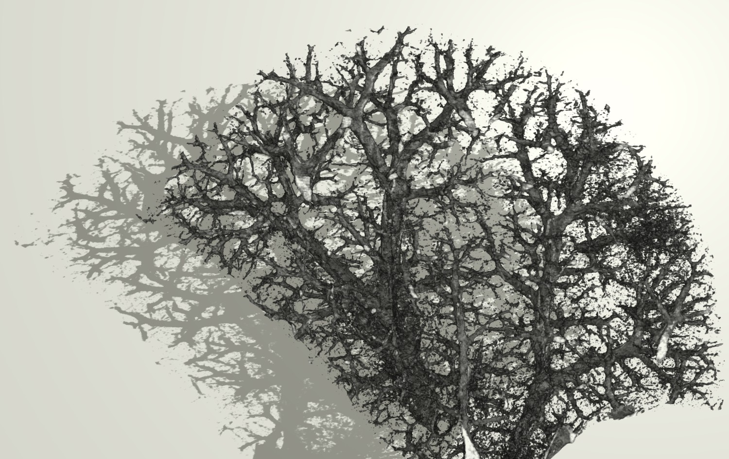
MINFLUX is the best bridge to structural biology that we currently have in light microscopy. It has the potential to add dynamic in vivo information at resolution levels that are not achievable in any other form. In an early paper, published in Nature Methods, the potential for imaging is exemplified when looking at nuclear pore complexes (NPCs) in vivo.
Since the installation of the first ever commercial MINFLUX instrument by Abberior two years ago in the EMBL Imaging Centre multiple projects were already successfully concluded:
For example, a study that was recently published in Science focuses on live-cell tracking of single fluorophores with nanometer spatial and millisecond temporal resolution in 2D and 3D.
Reference:
Deguchi T, Iwanski MK, Schentarra EM, Heidebrecht C, Schmidt L, Heck J, Weihs T, Schnorrenberg S, Hoess P, Liu S, Chevyreva V, Noh KM, Kapitein LC, Ries J. Direct observation of motor protein stepping in living cells using MINFLUX. Science. 2023 Mar 10;379(6636):1010-1015. doi: 10.1126/science.ade2676. Epub 2023 Mar 9. PMID: 36893247.
Another recent study looks at microtubule-binding proteins, distinguishing between those located in the internal lumen and on the external surface of the microtubule.
Reference:
van den Berg CM, Volkov VA, Schnorrenberg S, Huang Z, Stecker KE, Grigoriev I, Gilani S, Frikstad KM, Patzke S, Zimmermann T, Dogterom M, Akhmanova A. CSPP1 stabilizes growing microtubule ends and damaged lattices from the luminal side. J Cell Biol. 2023 Apr 3;222(4):e202208062. doi: 10.1083/jcb.202208062. Epub 2023 Feb 8. PMID: 36752787; PMCID: PMC9948759.
Another publication looks at self-blinking fluorescent probes for MINFLUX.
Reference:
Gerasimaitė RT, Bucevičius J, Kiszka KA, Schnorrenberg S, Kostiuk G, Koenen T, Lukinavičius G. Blinking Fluorescent Probes for Tubulin Nanoscopy in Living and Fixed Cells. ACS Chem Biol. 2021 Nov 19;16(11):2130-2136. doi: 10.1021/acschembio.1c00538. Epub 2021 Nov 4. PMID: 34734690; PMCID: PMC8609524.
We have now seen the amazing things MINFLUX is capable of visualising and its tracking resolution. What are some things researchers who would like to use MINFLUX should be aware of?
The data generation throughput can be limiting since the single localization events are collected sequentially. This allows for extremely good time resolutions when tracking a single molecule over time, but means that creating e.g. a 3D image dataset of a larger image field can take time. Also, single molecule tracks are of course only taken one at a time and not in parallel like in camera-based systems. This needs to be taken in account in the experimental planning. Given the achievable resolutions you must stringently optimize the label density, the label strategy and guarantee the stability of the sample over the measurement time. The sample preparation procedure is key and it requires a lot of expertise.
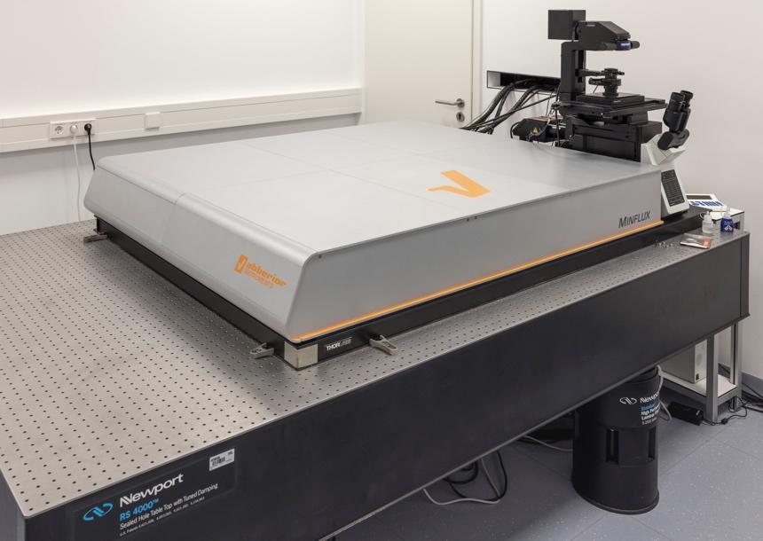
A photo of one of the two MINFLUX systems available for user projects at EMBL Imaging Centre.
What other services do you provide in your facility that would be useful in combination with this type of imaging?
We have two MINFLUX systems in the EMBL Imaging Centre and MINFLUX is offered as a full workflow, including sample preparation support and data analysis. With MINFLUX, we mainly work together with the user to optimize the workflow and service team staff actually performs the experiment. A lot of expertise is needed to run the MINFLUX system with respect to sample preparation, dyes, fiducial beads…. Every step has to be optimized to get a good result. The method is unforgiving. Only good samples lead to good results with the highest resolution possible and making optimal use of the method. MINFLUX data analysis is also still a developing field and the EMBL team provides data analysis expertise in their project pipeline to support the user.
We dedicate at least one week to every project. The first sessions are spent optimizing the project. To make the method more accessible, we have recently offered a training course in collaboration with Aberrior and we plan to offer such courses also in the future.
Learn more about MINFLUX at EMBL Imaging Centre here.
About MINFLUX at EMBL Imaging Centre.
How to apply:
MINFLUX is part of the Euro-BioImaging Proof-of-Concept study. The Proof-of-Concept study makes it possible to introduce exciting, new imaging technologies to our portfolio that were previously unavailable via our network. We are currently accepting applications to use these technologies at participating Nodes as part of the Proof-of-Concept study. Be part of this study - and contribute to community-wide continuous technological innovation!
All scientists, regardless of their affiliation, area of expertise or field of activity can benefit from Euro-BioImaging’s pan-European open access services. Potential users of these new technologies are encouraged to submit project proposals via our website. To do so, you can log in to access our application platform, choose the technology you want to use and the facility you wish to visit, then submit your proposal. All applications will be processed by the Euro-BioImaging Hub. As usual, users will benefit from advice and guidance by technical experts working at the Nodes, training opportunities, and data management services.
Meet Cristina Fernandez, Basque Resource for Electron Microscopy (BREM)
The Basque Resource for Electron Microscopy (BREM) provides access to high-end instrumentation and expertise in high-resolution cryo-electron microscopy (Cryo-EM) to national and international researchers,…