October 8, 2026
Euro-BioImaging at ‘Data Stewardship goes Germany – from going to growing’
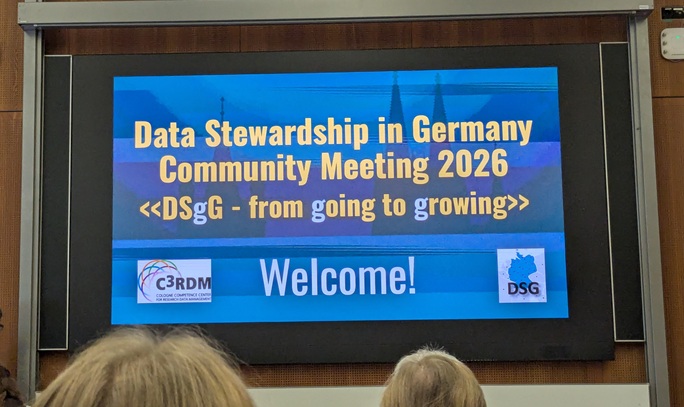
This September, Euro-BioImaging was part of the Data Stewardship goes Germany (DSgG) Community Meeting in Cologne. Under the theme “from going to…



The MorphoPHEN Master’s Program is an integrated and interdisciplinary Erasmus Mundus Joint Master, to train experts in morphological mouse phenotyping, to generate a positive impact of preclinical biomedicine and improve human translatability. It brings knowledge on the four main branches necessary to analyze mouse disease models morphologically: Anatomy and Embryology, Pathology, Imaging, and Deep Learning. In a stunning one-year curriculum that unfolds across four European countries: Italy, Portugal, Spain and Greece, students will be prepared to shape the future of preclinical research. Our MMMI Italian Node has contributed to developing the curriculum and several Node staff are involved in the Master’s Program as lecturers. We spoke with Sandra Albanese, of the Institute of Biostructures and Bioimaging- CNR, to learn more about the degree and the important role that preclinical imaging will play in it.
How did you become involved in the MorphoPHEN Master’s program?
Sandra Albanese: I became involved in the master's program by the University Federico II, Naples, Italy, to deliver lectures related to preclinical imaging and to conduct some practical activities in the laboratory.
What type of student profiles are you looking for?
Sandra Albanese: The profile of the master's student should have a propensity to deep in the field of preclinical research, particularly, those who wish to study models of human pathologies through the use of the most modern imaging techniques and sophisticated software.
Tell us about the Mouse Imaging, Module 2. How does it fit into the bigger picture of the curriculum?
Sandra Albanese: The Mouse Imaging Module 2 includes the construction of an integrated training program on morphological phenotyping of small animals, specifically on preclinical imaging of mouse models.
The aim is to provide insight into the physical principles and biomedical applications of imaging techniques while also highlighting the basics of animal care, handling, and preparation. Differences between various imaging methodologies (X-ray, CT, MRI, ultrasound, etc.), comparison of diagnostic possibilities among them, and different image processing will be described.
Why would you recommend this Master’s program?
Sandra Albanese: I recommend the master's program as it provides a broad view of the use of imaging techniques to analyze murine models of human pathology in a non-invasive manner.
Applications are being accepted until March 11, and scholarships are available! Don’t miss this opportunity to shape the future of preclinical research.
Visit the MorphoPHEN website: https://morphophen.eu
October 8, 2026
This September, Euro-BioImaging was part of the Data Stewardship goes Germany (DSgG) Community Meeting in Cologne. Under the theme “from going to…

October 7, 2026
On 28–29 September 2026, the growing French volume electron microscopy (vEM) community came together in Bordeaux for the first France VolumeEM Scientific Days.

October 7, 2026
Euro-BioImaging joined partners from across Europe on 29. & 30. September 2026 at the official kick-off meeting for STARDAST, a new Horizon Europe…