
March 30, 2026
Euro-BioImaging at the EOSC ESFRI meeting in Milan
Euro-BioImaging was delighted to attend the ESFRI/EOSC Policy Workshop on “EOSC and Research Infrastructures: Opportunities and Strategies” in Milan from March 15-16, represented by…

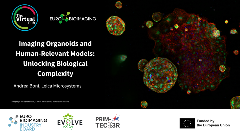
We will host the Special Edition Virtual Pub "Imaging Organoids" on Friday, January 30, from 1-3 pm CET. It will focus on methods used to image organoid systems. As part of this event, Karl Zhang, Center for Cellular Imaging, University of Gothenburg, Swedish NMI Node, will give a talk entitled "Optimizing Tissue Clearing Protocols and Imaging Techniques for Organoid study" (description below). Join us to hear this and other exciting presentations introducing a range of methods used to image organoid systems - from lung buds to mini-brains.
Title: Optimizing Tissue Clearing Protocols and Imaging Techniques for Organoid study
Presenter: Karl Zhang, Center for Cellular Imaging, University of Gothenburg, NMI Sweden Node
Organoids are advanced three-dimensional in vitro models that recapitulate key structural and functional characteristics of native tissues. However, their inherent architectural complexity poses significant challenges for high-resolution microscopy analysis. Accurate visualization of organoid architecture and dynamic biological processes depends not only on advanced imaging technologies but also on optimized sample preparation and clearing protocols.
In this presentation, I will explore various organoid sample clearing methods and demonstrate how different microscopy modalities—including confocal and light-sheet microscopy—can be strategically combined with appropriate preparation techniques to address specific research objectives in organoid studies.
March 30, 2026
Euro-BioImaging was delighted to attend the ESFRI/EOSC Policy Workshop on “EOSC and Research Infrastructures: Opportunities and Strategies” in Milan from March 15-16, represented by…
March 30, 2026
Euro-BioImaging was delighted to attend the Public Awareness & Engagement of Research Infrastructures (PAERI) conference, represented by External Communications Officer, Marianna Childress-Poli. This year’s…

March 26, 2026
The Madrid Advanced Microscopy Center (MAdMiC) is the first Euro-BioImaging Node in Madrid (Spain). It is formed through the collaboration and close work of…