This is a multi-sited Node comprising 8 imaging centers distributed in 4 cities (Torino, Pisa, Milan, and Naples) with the coordinator based in Torino. Three of our sites (Department of Molecular Biotechnology and Health Science - Torino, Clinical Physiology Institution CNR - Pisa, and Biostructures and Bioimages Institute CNR - Napoli) are equipped with photoacoustic technology.
Enzo Terreno, coordinator of the MMMI Italian Node (left) Martina Capozza, responsible for the activity at the Torino’s facility, at the PAI scanner (right)
Today we are talking about Photoacoustic Imaging. Please tell us a little bit about this type of imaging:
Photoacoustic imaging (PAI) is an emerging and powerful technology with a wide spectrum of applications in the field of in-vivo imaging. Briefly, Photoacoustic contrast originates from the absorption of a pulsed laser beam by a chromophore that causes a transient thermoelastic expansion of the surrounding tissues, responsible for the generation of acoustic waves that are detected by an Ultrasound (US) transducer and converted into images. That’s why PAI is considered as the “sound of the light.” PAI combines the high-contrast and the spectroscopic features of optical imaging in the near infrared region (NIR) with the high spatial-resolution and tissue penetration capability of ultrasound imaging. In vivo PAI can penetrate tissue from a few millimeters to centimeters of depth and gives an excellent spatial resolution of a few hundreds of microns attainable with and without using contrast agents. Finally, it is worth saying that this technique does not use ionizing radiation and can thus be considered safe.
Tell us a bit more about a specific project that was done in your facility using this technology? What scientific questions were you addressing?
The (preclinical) research activity on PAI in MMMI Italian Node is mainly focused to address the following topics:
- Exploit endogenous PAI contrast to assess vascular volume and oxygenation levels in tumours
- Design and validate innovative PAI probes, including systems for multimodal detection
- Pharmacokinetic for studying the distribution of labelled molecules of nanostructured materials.
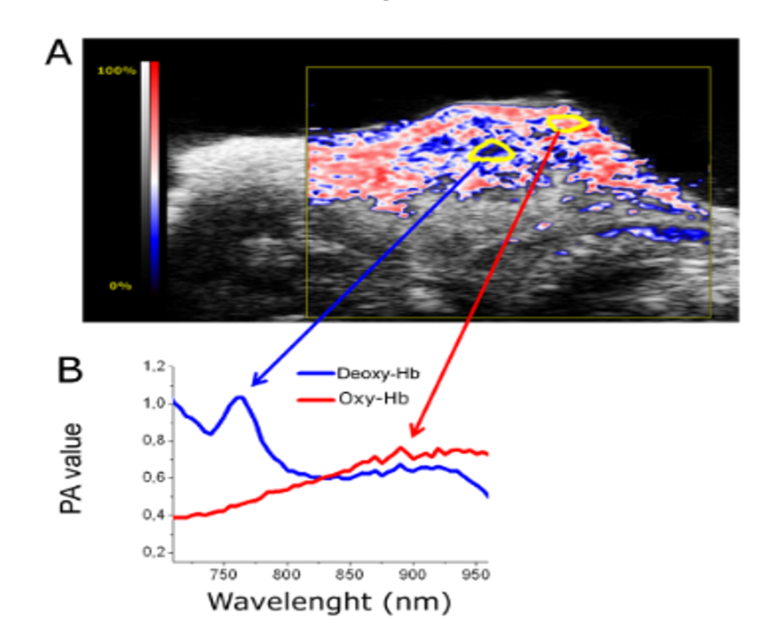
Here are two examples: 1) An MRI Method To Map Tumor Hypoxia Using Red Blood Cells Loaded with a pO2-Responsive Gd-Agent (https://pubs.acs.org/doi/10.1021/acsnano.5b02604). In this study, PAI has been used to non-invasively assess tumor vascular volume and tumor hypoxia, without the use of exogenous contrast agents. This allowed monitoring tumor growth and aggressiveness and validating MRI results on vascular volume and hypoxia.
(A) PA image of a subcutaneous mouse tumor showing ar-ea of hypoxia (blue) and normoxia (red). (B) PAI spectra of two regions (labelled in yellow) of the image showing the typical spectra of oxy-Hb (red) in the tumor rim and deoxy-Hb (blue) in the tumor core. (Di Gregorio et al., ACS Nano 2015, https://pubs.acs.org/doi/10.1021/acsnano.5b02604)
2) Photoacoustic imaging of integrin-overexpressing tumors using a novel ICG-based contrast agent in mice. (https://pubmed.ncbi.nlm.nih.gov/30105205/). In this study, a novel integrin-targeting photoacoustic probe was synthesized to combine the good PAI properties of ICG and the favourable αvβ3-binding capabilities of a cyclic RGD peptidomimetic moiety previously used for fluorescence imaging.
A) Representative PA images of a mouse tumor region acquired 15 min, 60 min and 24 h post in-jection of the PA probe and spectrally unmixed. The ROIs used for data analysis are indicated in red; B) PA signal of tumor regions at different timepoint after injection (n = 5/group). *P < 0.01,**P < 0.001, ns P > 0.05, 2-way ANOVA re-peated measures followed by Bonferroni post-hoc test. (Capozzo et al., PhotoAcoustics, 2018. https://pubmed.ncbi.nlm.nih.gov/30105205/)
Why is this technology best suited to address that question?
PAI has the important strength, unique compared to in vivo imaging technologies, to provide a quantita-tive assessment of tissue perfusion and oxygenation without using exogenous probes. Furthermore, the development of specific and innovative probes is fundamental for extending the poten-tial of this technology.
What other services do you provide in your facility that would be useful in combination with this type of imaging?
The MMMI Italian Node is equipped with all the state-of-the-art in vivo imaging technologies. Among them, high frequency ultrasound is naturally combined with PAI, and all the PAI scanners in the Node are fully integrated with an Ultrasound imager. In principle, PAI can be combined with optical and MR scan-ners, though efforts are required for allowing the co-localization of the two images.
Why should scientists choose your facility for using this technology?
The three Centers of the MMMI Italian Node equipped with PAI technology have also a strong compe-tence and active collaborations for the design of innovative PA probes. Furthermore, MMMI Italian Node has a longstanding and strong expertise in all the molecular and clinical imaging technologies represented in Euro-BioImaging portfolio (MRI, nuclear medicine, optical imaging, ultrasound), thus allowing the exe-cution of validation and comparative investigations on site. And with the current travel restrictions in place, it is worthwhile to note that all of our imaging services are provided remotely. We provide a turn-key solution, elaborated in close collaboration with the user, but there is no need for the user to be on site.
Learn more about how remote experiments are run at the MMMI Italian Node.
How to apply:
In July 2020, Euro-BioImaging launched a Proof-of-Concept study - in collaboration with its Nodes - making it possible to access six previously unavailable imaging technologies. This article – the fourth in a series - features interviews with Euro-BioImaging Node staff, shedding light on some potential applications of these technologies - and providing advice on how to get the most out of them. We are currently accepting applications to use these technologies as part of the Proof-of-Concept study.
All scientists, regardless of their affiliation, area of expertise or field of activity can benefit from Euro-BioImaging’s pan-European open access services.Potential users of these new technologies are encouraged to submit project proposals via our website. To do so, you can Login to access our application platform, choose the technology you want to use and the facility you wish to visit, then submit your proposal. All applications will be processed by the Euro-BioImaging Hub. As usual, users will benefit from advice and guidance by technical experts working at the Nodes, training opportunities, and data management services.
A Multiplexed Vision: Highlights from the Special Edition Virtual Pub on Spatial Transcriptomics
On June 12th, Euro-BioImaging hosted its latest Special Edition Virtual Pub, bringing together over 200 researchers and technology developers working at the cutting…
Cellular Imaging Hungary Node expands expertise in advanced neurophotonics
The Euro-BioImaging Cellular Imaging Hungary Node has expanded its service portfolio with the addition of the BrainVisionCenter (BVC) in Budapest. Following a successful…
The UK Euro-BioImaging Node expands from seven to thirteen sites!
We’re delighted to announce that the UK Euro-BioImaging Node is expanding, growing from seven to thirteen sites following a successful upgrade application and…