
March 30, 2026
Euro-BioImaging at the EOSC ESFRI meeting in Milan
Euro-BioImaging was delighted to attend the ESFRI/EOSC Policy Workshop on “EOSC and Research Infrastructures: Opportunities and Strategies” in Milan from March 15-16, represented by…

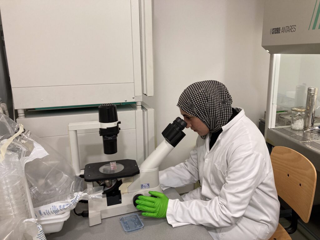
Colorectal cancer deaths are rising among young adults. Rabe’ah Almahassneh, a PhD student from the University of Valencia who is developing her research work at the Centro de Investigacion Principe Felipe, is investigating mutations in the APC protein (Adenomatous Polyposis Coli), found in 85% of colorectal cancer cases. Her research project aims to understand how these mutations impact cell adhesion and migration, potentially driving cancer in young patients. To explore this, Rabe’ah engineered APC mutations and tested them in 2D and 3D spheroid models of colorectal cancer cells. Using these models and live-cell confocal imaging, her team found altered focal adhesions in mutant cells, a clue to colorectal cancer progression. To deepen her study, and supported by France-BioImaging, Rabe’ah turned to the IGBMC photonic microscopy platform in Strasbourg. This platform offers open access to imaging technologies and expertise via Euro-BioImaging as part of France-BioImaging’s Alsace Node.
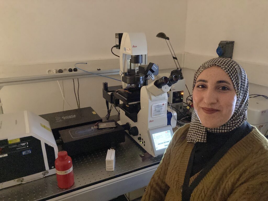
During her stay, Rabe’ah employed total internal reflection microscopy (TIRF), a powerful technique enabling non-invasive, high-resolution imaging of live cells. This technology allows her to analyze focal adhesion dynamics with unprecedented detail. “TIRF is essential to explore focal adhesion assembly and disassembly,” Rabe’ah explains. “The Strasbourg platform provides everything I need to conduct my experiments, from wet lab spaces to image analysis support”, says Rabe’ah, who hopes to bring this technology to her home lab.
The IGBMC platform exemplified international scientific collaboration as the IGBMC team provided state-of-the-art tools, training, and support, enabling breakthroughs that may illuminate the molecular drivers of colorectal cancer. Rabe’ah benefited from the strong involvement of two engineers of the platform: Erwan Grandgirard from the beginning of the discussion and first steps of the collaboration organization to the microscopy training, everyday support and image analysis, and Yves Lutz, all time assistance for the wet lab space especially cell culture, reagents as well as help for accommodation conditions.
“We welcome nearly 200 users from the IGBMC and outside annually,” says Yves Lutz. “Our guest house makes mid-to-long-term projects, like Rabe’ah’s, seamless for international researchers.”
With advanced imaging setups, dedicated training, hands-on support and wet lab space, the facility is a cornerstone for research that bridges molecular insights with real-world impact.
Rabe’ah’s work here exemplifies how access to such technologies can push the boundaries of understanding, offering hope for tackling colorectal cancer more effectively.
Article written by Daniela Aviles Huerta
“I was fortunate to gain access grant to the France BioImaging platform at the Alsace node (IGBMC, Strasbourg) to do experiments using Total Internal Reflection Fluorescence (TIRF). Throughout my time at the facility, I received exceptional support from the team. Their expertise and assistance greatly enhanced the efficiency and productivity of my experience. The IGBMC imaging platform offered an excellent environment, equipped with all the necessary reagents and materials for my experiments.
I acquired skills in TIRF, super-resolution microscopy, and Imaris software, which played a crucial role in the successful completion of my project. This experience yielded promising results that would not have been achievable without the team's support and the outstanding infrastructure provided by France-BioImaging.
I sincerely appreciate this opportunity and would strongly recommend the platform to fellow researchers seeking high-quality imaging resources and expert assistance.”
- Rabe’ah Almahassneh, PhD student, University of Valencia
March 30, 2026
Euro-BioImaging was delighted to attend the ESFRI/EOSC Policy Workshop on “EOSC and Research Infrastructures: Opportunities and Strategies” in Milan from March 15-16, represented by…
March 30, 2026
Euro-BioImaging was delighted to attend the Public Awareness & Engagement of Research Infrastructures (PAERI) conference, represented by External Communications Officer, Marianna Childress-Poli. This year’s…

March 26, 2026
The Madrid Advanced Microscopy Center (MAdMiC) is the first Euro-BioImaging Node in Madrid (Spain). It is formed through the collaboration and close work of…