April 30, 2026
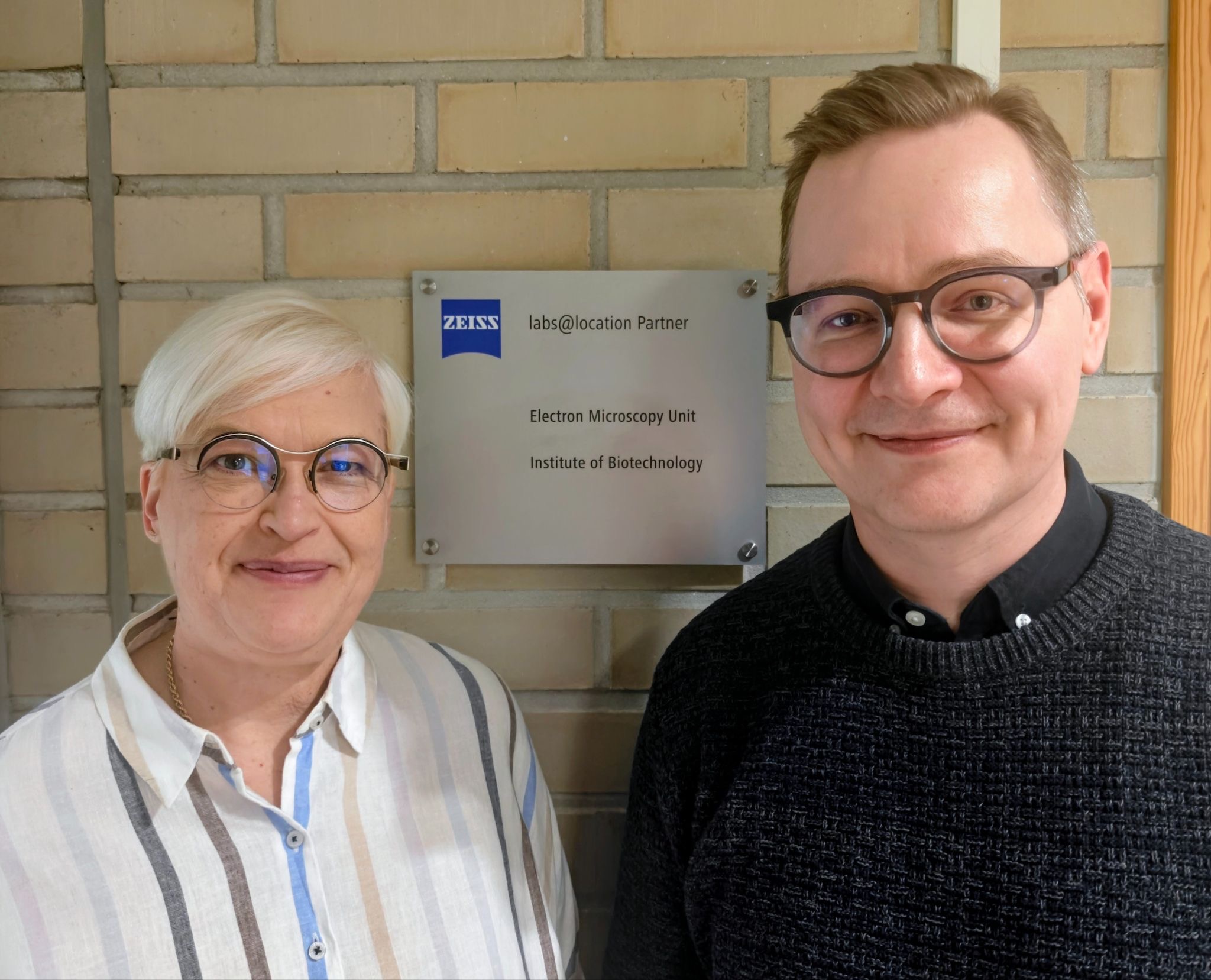
Euro-BioImaging facility in Helsinki selected to be first Zeiss Labs@Location site in Finland!
The Electron Microscopy Unit (EMBI) provides biological electron microscopy services at the University of Helsinki. Led by Research…

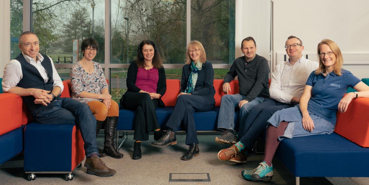
Spatial Transcriptomics is a useful approach for understanding tissue architecture and for understanding the molecular basis of health and disease. The Bioscience Technology Facility at the University of York, part of our UK Node, has experience with this exciting technique and is now accepting applications for spatial transcriptomics projects as part of the Euro-BioImaging Proof-of-Concept study. We spoke with Peter O’Toole, Director of the Bioscience Technology Facility, to find out more about this approach and what it can be used for.
We are today talking about Spatial Transcriptomics. Please provide a short summary of this approach and list some applications:
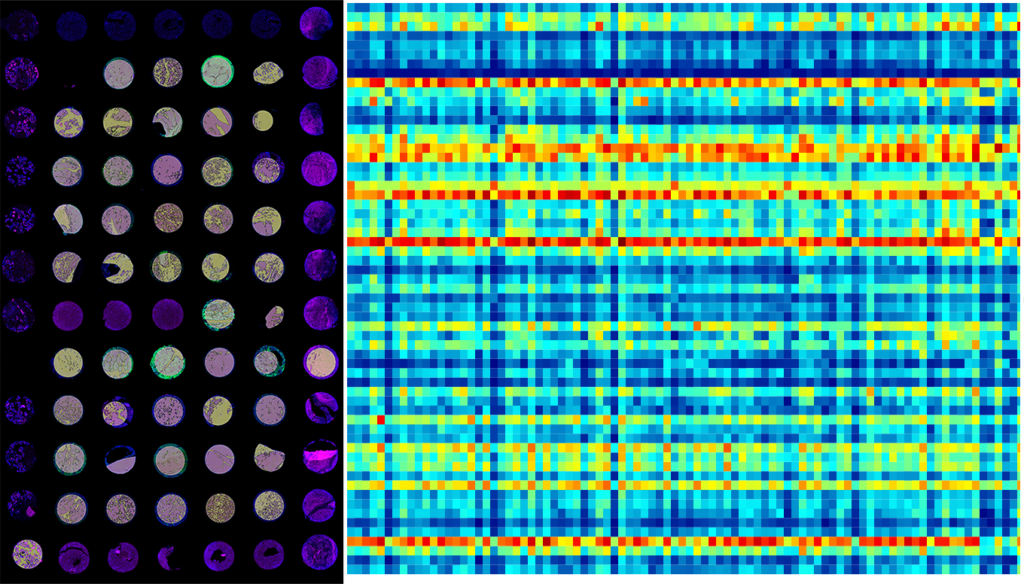
Spatial transcriptomics is a specific form of spatial ‘omics, to look at the spatial localisation of thousands of expressed RNA transcripts. Depending on the technology, localisation precision can be at the tissue level (tumor vs Tumor Microenvironment (TME)) or at the single cell level with subcellular detail, in Formalin-Fixed Paraffin-Embedded (FFPE) or cryo sections. RNA transcript species can be determined using a robust targeted approach that can be a customised or non-targeted approach for exploratory analysis. Each technique has its own advantages, such as being able to look at a whole tissue at speed, through to sub-sections of tissue with high resolution. Similar approaches are available for protein expression for 50+ proteins (Proteomics).
Transcriptomics can be useful for identifying gene expression differences within a single biopsy sample for cancer or neural studies. Proteomics can be used to profile protein expression to phenotype the immune response around an infected area of tissue. The list of applications is very varied. Although most are still focused on human and mouse tissues, some approaches are now also enabling more diverse sample types including plant materials.
Tell us a bit more about a specific project that was done in your facility using this approach? What scientific questions were you addressing?
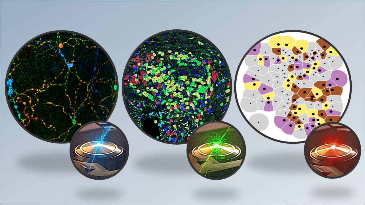
With the Kaye group at University of York, we have been using spatial ’omics looking at immune response through detection of proteins and RNA localisation for studying how the immune system reacts to leishmanaiasis infections.
References
We have extended this collaboration with the Kaye group and the Heeren group in Maastricht University and linking the spatial phenotyping/transcriptomics with spatial lipidomics, really using the power of all of these together to study sequential tissue sections.
References
What are some advantages of this technique that make it suited to addressing this type of question?
Flow cytometry is a great way for phenotyping cells, as is sequencing approaches for looking at RNA expression levels within tissues, but these do not typically give any spatial context. It is the heterogeneity that is needed to understand how the tumours are interacting with their local environment, and this can dramatically vary from site to site within a singular tissue section. The spatial awareness now enables us to embrace this heterogeneity and identify precisely how the cells are being manipulated or responding to challenges.
What other services do you provide in your facility that would be useful in combination with this type of imaging?
Many of these samples also require imaging on more routine confocal microscopes. We take full field of view images using the Zeiss AxioScan, and higher resolution tiled images of tissue sections using either the confocal microscopes or ELYRA7 using the Apotome mode. Naturally, we have already used the Euro-BioImaging Maastricht Node ourselves as part of the collaboration for matching up our spatial ’omics approaches with their spatial lipidomic approaches, and doing similar work our in house Waters Synapt for spatial metabolomic studies of sequential samples. It also goes without saying that many of these groups start these studies without spatial awareness using flow cytometry, which is also supported within our own facility with over 9 cytometers on hand.
We can accommodate up to 5 spatial transcriptomics projects per year at our facility. If you are interested in working collaboratively with us, please discuss with us at York in advance so that we can check the sample suitability. We can provide support for human and animal tissues. We can then help with pricing, sample number required, and practicalities required.
How to apply:
Spatial transcriptomics is part of the Euro-BioImaging Proof-of-Concept study. The Proof-of-Concept study makes it possible to introduce exciting, new imaging technologies to our portfolio that were previously unavailable via our network. We are currently accepting applications to use these technologies at participating Nodes as part of the Proof-of-Concept study. Be part of this study - and contribute to community-wide continuous technological innovation!
All scientists, regardless of their affiliation, area of expertise or field of activity can benefit from Euro-BioImaging’s pan-European open access services. Potential users of these new technologies are encouraged to submit project proposals via our website. To do so, you can log in to access our application platform, choose the technology you want to use and the facility you wish to visit, then submit your proposal. All applications will be processed by the Euro-BioImaging Hub. As usual, users will benefit from advice and guidance by technical experts working at the Nodes, training opportunities, and data management services.
For more information: info@eurobioimaging.eu
April 30, 2026
The Electron Microscopy Unit (EMBI) provides biological electron microscopy services at the University of Helsinki. Led by Research…
April 29, 2026
We are delighted to announce that a new Node has joined Euro-BioImaging after stringent review by our Scientific Advisory Board and approval by the…
April 29, 2026
A new publication from the Global BioImaging community sheds light on the career paths, recognition, and working conditions of imaging…