Special Edition Virtual Pub: Spatial Proteomics


Published November 11, 2026

Proteins, Pixels and a Pint: Highlights from the Spatial Proteomics Virtual Pub

Last orders were called on the transcripts – this time, the proteins got their round. On October 2nd, Euro-BioImaging hosted its latest Special Edition Virtual Pub, this time dedicated to spatial proteomics. As promised at the end of our spatial transcriptomics edition, we turned our attention to the spatial distribution of proteins. The line-up ran from cloud-based AI analysis and in situ protein–protein interaction readouts to mass spectrometry at single-region resolution and national-scale visions for joining microscopy and omics. If there was one theme on tap, it was this: spatial proteomics is no longer just about making pretty multiplexed images, but about making them quantitative, scalable and genuinely useful for biology and the clinic.

Article by Sanjana Singh, Scientific Project Manager

An afternoon for the spatial biology community

The Special Edition Virtual Pub format once again brought together imaging scientists, core facility staff, technology developers and industry representatives. Five speakers took the stage, two from academic facilities and infrastructures and three from companies building the next generation of spatial tools. Grab a drink – here is what they served up.

Visit the playlist

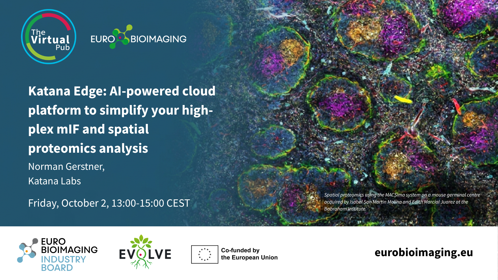
Norman Gerstner, Katana Labs: Katana Edge: AI-powered cloud platform to simplify your high-plex mIF and spatial proteomics analysis

Poster

Norman opened the session by tackling every facility's favourite headache: what to do with enormous high-plex datasets. He introduced Katana Edge, a cloud-native platform that lets teams analyze ultra-high-plex whole-slide images and TMAs directly in the browser – no in-house compute cluster required. AI workflows handle cell and tissue segmentation, phenotyping and clustering across mIF, spatial proteomics, spatial transcriptomics, FISH and IHC. Bonus points for the LLM-powered option to simply ask your data questions.

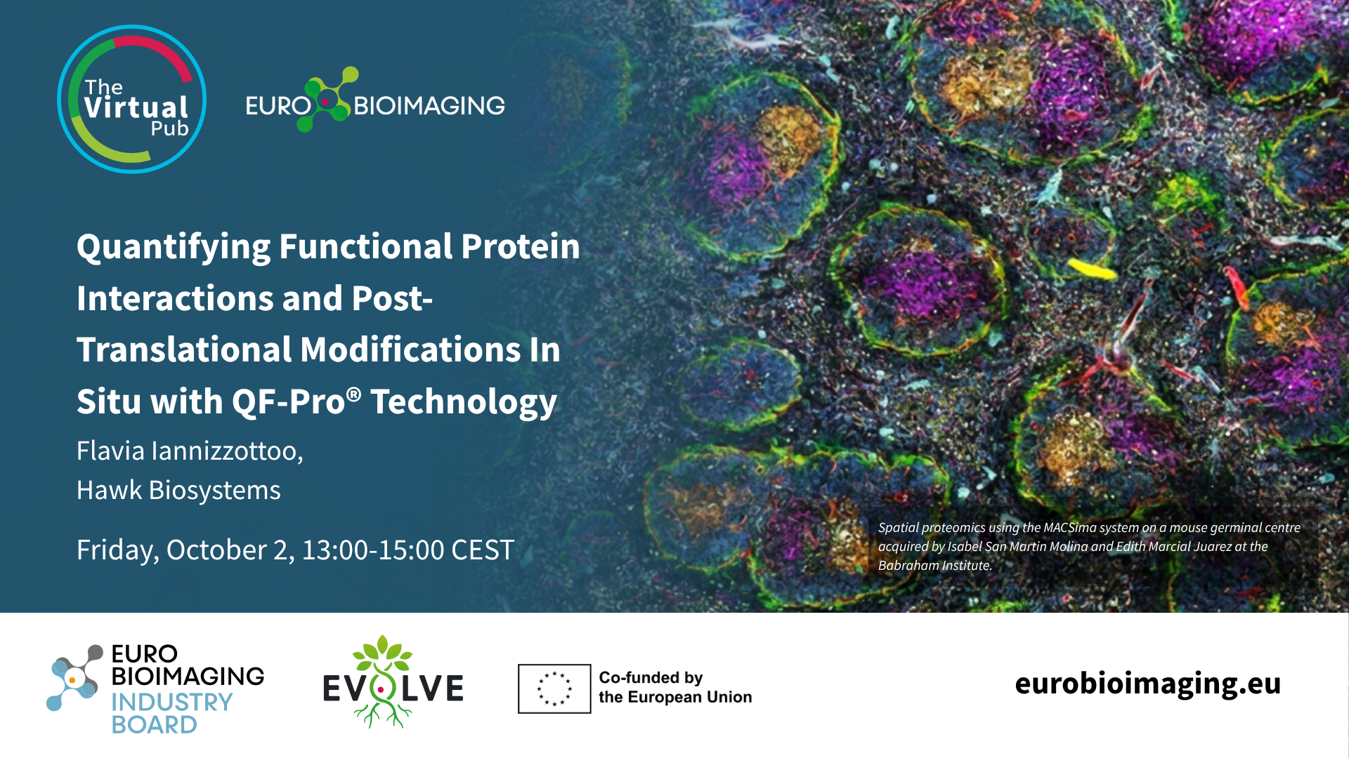
Flavia Iannizzotto, Hawk Biosystems: Quantifying Functional Protein Interactions and Post-Translational Modifications In Situ with QF-Pro® Technology

Poster

Knowing where a protein sits is one thing; knowing whether it is actually doing anything is another. Flavia showed how QF-Pro® adds that functional layer, using amplified FRET-FLIM to quantify protein–protein interactions and post-translational modification states in fixed cells and FFPE tissue. The result is a single QF-Pro® Score that reflects how engaged two partners really are. Because it runs independently of other omics workflows, it can be layered onto existing spatial pipelines – with an eye on future companion diagnostics.

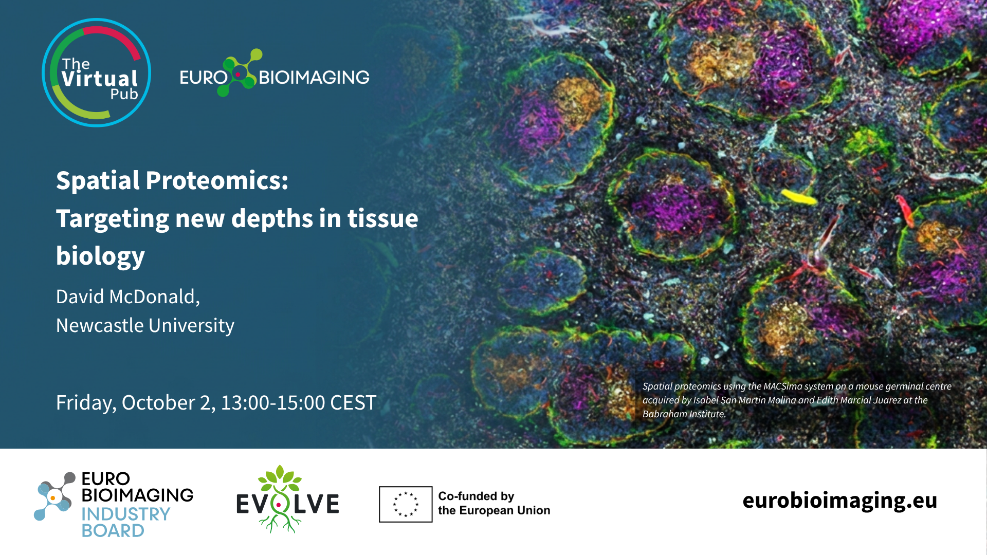
David McDonald, Newcastle University: Spatial Proteomics: Targeting new depths in tissue biology

Virtual Pub Special Edition Poster

David set a course for the final frontier – quite literally, opening with a Star Trek-inspired mission statement "to boldly go where no antibody has gone before." Back on Earth, he asked an honest question: if spatial proteomics relies on fairly simple tools and common skills, why do so many labs struggle to make it work? He shared how Newcastle's facility-led approach helps users navigate the time, cost and complexity, delivering results across a wide range of tissues, targets and diseases.

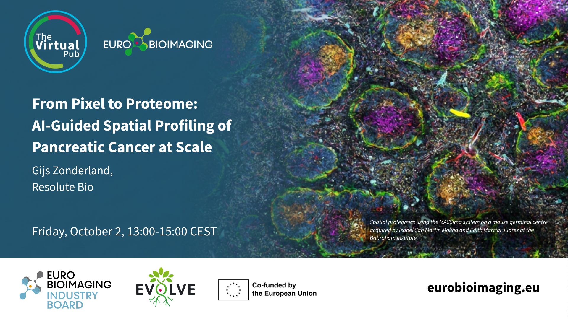
Gijs Zonderland, Resolute Bio: From Pixel to Proteome: AI-Guided Spatial Profiling of Pancreatic Cancer at Scale

Resolute Bio poster

Gijs took us from pixel to proteome by coupling AI-guided pathology with Deep Visual Proteomics. Foundation models first located precursor lesions in pancreatic cancer tissue; laser microdissection and mass spectrometry then quantified more than 9,000 proteins from around 100 cells. The headline finding: proteome-level reprogramming happens beforethe tissue looks any different under the microscope. Scaled up as tileDVP, the approach generated over 2,500 proteomic measurements across 55 FFPE blocks, building an atlas from earliest precursor to metastatic disease.

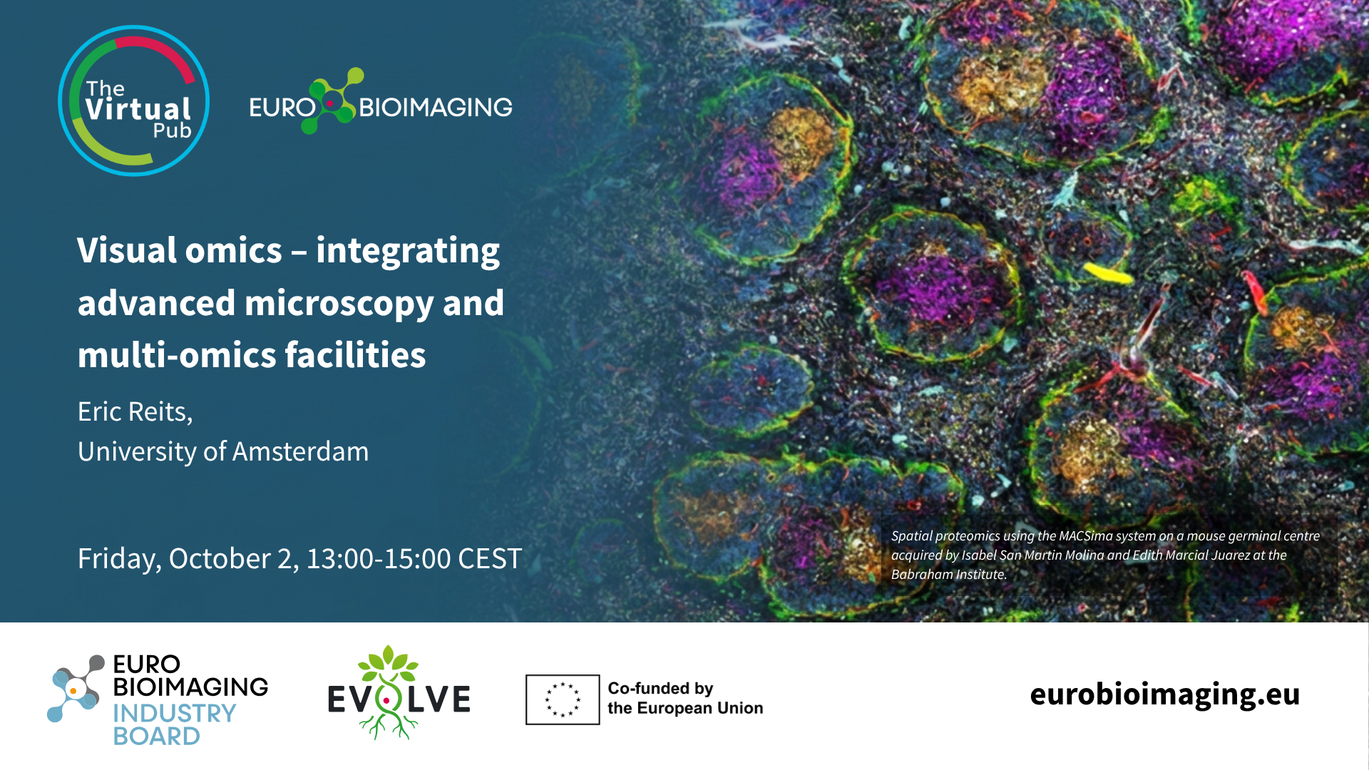
Eric Reits, University of Amsterdam: Visual omics – integrating advanced microscopy and multi-omics facilities

Poster

Eric closed the session with the big picture. He presented Visual Omics, the next-generation integration of NL-BioImaging (home of the Advanced NL microscopy Euro-BioImaging Node) and the Netherlands X-omics Initiative. The goal: move beyond static snapshots to "record" the molecular history of living cells by linking microscopy and omics facilities within and between institutes. He outlined the challenges to tackle at national level, from smart microscopy that automatically picks out cells for omics analysis to truly cross-modal data integration.

Thank you and don't miss the next event!

A big thank you to all our speakers and attendees for making this Special Edition such a success. Keep an eye on our events page – the next round is already being poured. Join us for the Special Edition Virtual Pub "Imaging Micro- and Nanoplastics." Cheers!


More news from Euro-BioImaging