June 3, 2026
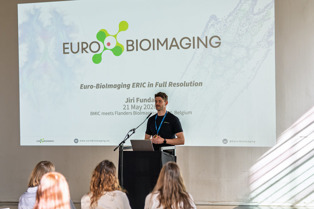
BMIC meets Flanders BioImaging
In May 2026, the Belgian Molecular Imaging Community (BMIC) and Flanders BioImaging joined forces in Ghent for a two-day community event that brought…

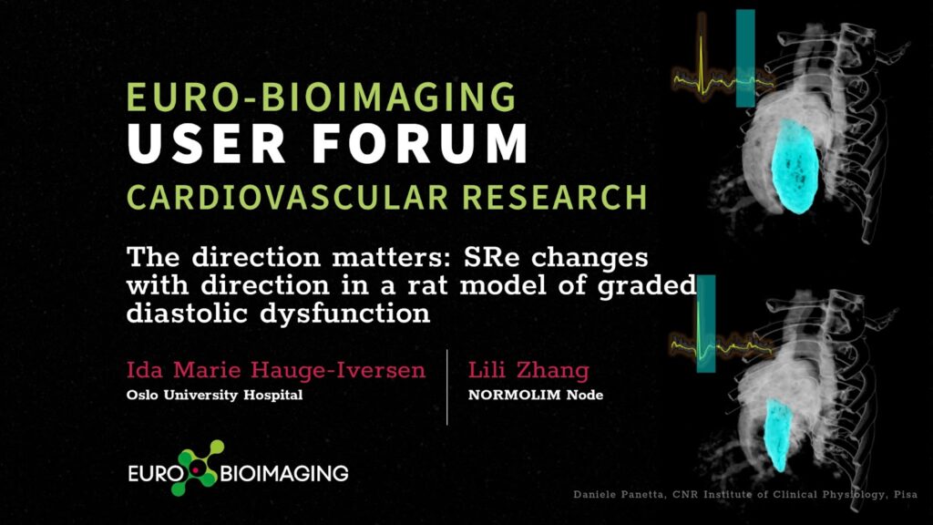
Euro-BioImaging is organizing an online User Forum on Tuesday, March 21, 2023, from 14:00-17:00 CET. The topic is “Cardiovascular research.” This event will highlight the importance of cutting-edge imaging technologies in support of cardiovascular health, disease, diagnostics and the development of therapies. We will showcase the specific expertise available at our Nodes across Europe through case studies presented in tandem with the research community.
At this event, Ida Marie Hauge-Iversen, Oslo University Hospital, will explain how tissue phase mapping (TPM) Cardiac magnetic resonance (CMR) which she performed at the NORMOLIM Node, can be used to assess bidirectional Myocardial strain rate in the early filling phase (SRe), and potentially bring us one step closer to understanding the diastolic function of the heart.
Hear this talk and others like it on March 21 at the Euro-BioImaging User Forum: Cardiovascular Research.
Abstract:
Diastolic dysfunction (DD) is an increasing public health problem, where important underlying mechanisms remain unknown. Accurate measures of DD in animal models are crucial for mechanistic research. Myocardial strain rate in the early filling phase (SRe) is closely associated with diastolic function. Cardiac magnetic resonance (CMR) offers unparalleled ability to quantify myocardial deformation rates in any direction. In this study, we utilized tissue phase mapping (TPM) CMR to assess bidirectional SRe in a rat model of graded DD. The aim was to get better knowledge of how DD affects SRe in different directions.
Graded DD was induced in rats by aortic banding (AB), where o-rings with an inner diameter of either 1.3 (n=8) or 1.5mm (n=10) were placed around the ascending aorta. A group with sham-operated rats (n=10) served as control. MRI was performed 20 weeks post-operation, when left ventricle (LV) hypertrophy and myocardial fibrosis were allowed to develop. CMR with TPM and CINE was performed using a 9.4T preclinical CMR system, before tissue was harvested. TPM included a long-axis four-chamber view (for longitudinal SR) and a mid-LV short-axis view (for circumferential SR). CINE included a short-axis stack of the whole LV and left atrium (LA). SRe was calculated from TPM using Matlab. Ex vivo tissue was weighed and analyzed by ddPCR. p-values were found using Kruskal-Wallis with multiple comparison test.
Both AB groups showed signs of DD with increased LV mass, increased BNP expression and preserved EF. In addition, the 1.3 group exhibited higher collagen expression, lung weight and LA volume than the 1.5 group, indicating more severe DD. Longitudinal SRe was lower (p=0.01) in the 1.3 group compared to control, and exhibited a negative correlation to LA volume across all three groups. Circumferential SRe was higher (p=0.01) in the 1.3 group compared to control and showed a positive correlation to LA volume.
We found that with increasing grade of DD, longitudinal SRe is reduced, whereas, interestingly, circumferential SRe is increased. This may indicate that improved circumferential SRe compensate for the loss in longitudinal SRe. Our results demonstrate the importance of the direction in which myocardial deformation rate is measured. Further investigation of this mechanism can potentially bring us one step towards understanding the diastolic function of the heart.
June 3, 2026
In May 2026, the Belgian Molecular Imaging Community (BMIC) and Flanders BioImaging joined forces in Ghent for a two-day community event that brought…

June 1, 2026
The EOSC Association has adopted a new position paper calling for a distinct Work Programme-based Partnership for EOSC in FP10. Euro-BioImaging welcomes this…

May 29, 2026
Spatial transcriptomics is rapidly transforming the way researchers study complex biological systems. To explore the latest developments in this fast-moving field, Euro-BioImaging is…