Join the Special Edition Virtual Pub, “Innovation in Imaging,” where we will discuss some of the issues that innovators should be aware of and hear use cases from our network about successful tech transfer and translation from the lab.
When: September 27, 2024, from 13:00-15:30 CEST
Where: Online
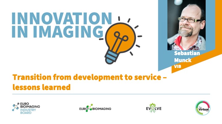
At this event, Sebastian Munck (VIB), will explain how establishing workflows and committing staff time to innovative/development activities can be beneficial for end-users, in a presentation entitled, “Transition from development to service – lessons learned.”
Transition from development to service – lessons learned
Sebastian Munck, VIB
Abstract
Core facilities are vital for providing services to departments and stakeholders, but without innovation, they risk stagnation and failure to advance science.
Core facilities are often based at discovery-based life science institutes, where development is a distinct activity and where a development project within a core facility is a temporary endeavour to produce a unique product, service, or result, turning a challenge into a solution. These projects vary in complexity, ranging from complete new imaging methods to protocol optimizations and technology adaptation.
Establishing workflows and committing staff time to innovative/development activities that benefit the majority of users is essential for creating scalable development projects for long-term service implementation. A road map with go-and-no-go decisions and milestones can make this process transparent.
Building Skills Across Europe: EVOLVE Supports the Second Edition of the Distributed Image Analysis Training Course
The second edition of the “Introduction to Image Analysis with Python for Life Scientists” course marked another successful milestone in the development of Euro-BioImaging’s distributed training model.
Strengthening research-industry partnerships: results from the EVOLVE industry job shadowing pilot
The EVOLVE project, supported by European Union funding, recently completed a pilot programme designed to deepen the relationships between academic research infrastructures…
Imaging-Driven Neuroscience, Responsible Research, and the Power of Advocacy: Highlights from FENS Forum 2026
Euro-BioImaging was pleased to participate in the FENS Forum 2026, Europe’s largest international gathering dedicated to advancing neuroscience (>8000 participants). Throughout…