
March 30, 2026
Euro-BioImaging at the EOSC ESFRI meeting in Milan
Euro-BioImaging was delighted to attend the ESFRI/EOSC Policy Workshop on “EOSC and Research Infrastructures: Opportunities and Strategies” in Milan from March 15-16, represented by…

Optical Coherence Tomography (OCT) is a label-free technique that has emerged as an excellent tool for monitoring the structure and function of organoids. Nevertheless, organoids are usually too opaque, limiting the application.
In their work on acoustofluidic platforms for trapping and rotational manipulation of biological samples for tomography, Dr. Mia Kvåle Løvmo and Prof. Monika Ritsch-Marte, from Institute of Biomedical Physics, Medical University of Innsbruck, investigate how to overcome this issue. They thought that one possible solution would be having multi-angle views of the sample. Together with their collaborators at the Medical University of Vienna, Prof. Wolfgang Drexler and Dr. Shiyu Deng, they launched an exciting project to combine for the first time acoustic trapping and rotation with OCT. Access to OCT technology, along with related services and expertise, is provided by the Austrian BioImaging/CMI Node.
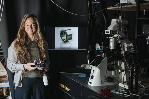
The acoustofluidic device consists of a 3D-printed chamber and transducers that can be inserted into an OCT imaging system to levitate and reorient suspended zebrafish larvae and tumor spheroids in a controlled and reproducible manner for multi-angle-OCT.
Importantly, the acoustic trapping proved to trap the samples stably which was suitable for the scanning-based OCT acquisition carried out by Dr. Deng.
Animated illustration of acoustic manipulation of levitated zebrafish embryo. Acoustic standing waves (green) are controlled to trap and rotate the sample inside the fluid-filled chamber. Transient rotation allows for optical imaging (red beam) from multiple directions, e.g., for multi-angle high-speed OCM through the bottom cover glass of the 3D printed octagon frame (black) (Blender rendering by Kvåle Løvmo)
The price to pay when using a non-contact method to trap and rotate a sample is that its positions and orientations are not known a priori. Overcoming this challenge was the other crucial step toward enabling accurate reconstruction from multi-angle OCT data. Dr. Simon Moser, also from the Institute of Biomedical Physics at the Medical University of Innsbruck, addressed this problem by developing a model-based reconstruction algorithm that allows the physically consistent fusion of multi-angle data acquired at a priori unknown object angles. Despite some current limitations, which will be addressed in future work, the method already succeeds in providing volumetric information with enhanced penetration depth—in a non-contact manner, and without requiring moving mechanical parts or supporting scaffolds. The authors believe the novel and non-invasive technique will be particularly important for long-term studies of developing organoids.
Read more: Ultrasound-induced reorientation for multi-angle optical coherence tomography - http://dx.doi.org/10.1038/s41467-024-46506-2
Austrian BioImaging/CMI is a multi-sited, multimodality Node that spans the full spectrum of biological and biomedical imaging – from cryo-electron and advanced light microscopy to preclinical modalities such as microCT, microMRI, microPET. The Node unites eight leading Austrian institutions and universities, which together provide a broad portfolio of services for organic materials, biomedical model organisms and large animals.
Through Austrian BioImaging/CMI users gain access to numerous multimodality imaging pipelines and specialized support services, including data and image analysis. Imaging technologies can be flexibly combined into tailored workflows or applied as stand-alone methods, depending on the specific research question. Covering the complete resolution range relevant for biological and preclinical studies, the imaging techniques deliver complementary insights into structure, function, dynamics and chemical composition.
With more than 30 imaging modalities available, Austrian BioImaging/CMI enables both in vitro, in vivo and ex vivo imaging, as well as molecular analysis. The Node also offers access to unique expertise in specialized techniques, such as in Optical Coherence Tomography (OCT), X-Ray Fluorescence (XRF) and Mass Spectroscopy Imaging, High-Resolution Episcopic Microscopy (HREM), or high-throughput plant phenotyping facility. A particular strength of Austrian BioImaging/CMI lies in the development of advanced multimodality workflows at the forefront of correlated imaging, often combining more than two imaging modalities into cutting-edge solutions for complex research challenges.
March 30, 2026
Euro-BioImaging was delighted to attend the ESFRI/EOSC Policy Workshop on “EOSC and Research Infrastructures: Opportunities and Strategies” in Milan from March 15-16, represented by…
March 30, 2026
Euro-BioImaging was delighted to attend the Public Awareness & Engagement of Research Infrastructures (PAERI) conference, represented by External Communications Officer, Marianna Childress-Poli. This year’s…

March 26, 2026
The Madrid Advanced Microscopy Center (MAdMiC) is the first Euro-BioImaging Node in Madrid (Spain). It is formed through the collaboration and close work of…