
October 12, 2026
EVOLVE Mentoring Programme 2026: Building connections across the imaging community
The second edition of the EVOLVE Mentoring Programme has come to a close, marking the end of several months of mentoring, knowledge exchange…



Therapies that target rapidly dividing cells have transformed the treatment of many cancers. Yet some promising therapies lose their effectiveness over time, allowing tumors to keep growing. Postdoctoral researcher Petra Hyroššová and PhD candidate Pavel Jakoubě, from the Institute of Biotechnology of the Czech Academy of Sciences, are investigating why this happens. By studying both the cancer cells and the healthy cells that surround them, they hope to better understand how tumours adapt to treatment. Thanks to canSERV funding, they accessed multiplex spatial phenotyping and confocal microscopy at Imaging Methods Core Facility at BIOCEV (IMCF), part of Euro-BioImaging's Advanced Light & Electron Microscopy Prague Node, to explore the tumour microenvironment and its response to therapy.
Although cancer therapies are designed to target tumour cells, tumours themselves are complex ecosystems made up of many different cell types, including blood vessels, immune cells and connective tissue. Increasing evidence suggests that these healthy cells can also influence how tumours respond to treatment. Petra's research focuses on endothelial cells, which line the tumour blood vessels and are among the first cells to encounter drugs circulating through the bloodstream. She wants to understand whether changes in these cells help explain why therapies become less effective over time.
Meanwhile, Pavel is investigating how the cancer cells themselves continue to proliferate despite the therapy.
"We are looking at tumours in vivo, where many different cell types interact," says Petra. "If you want to study this in a comprehensive way, you need a comprehensive technique. Multiplex spatial phenotyping allowed us to start broad and then narrow it down, studying our cells in their natural context."
"The spatial proteomics workflow was technically challenging. The IMCF team helped us enormously with optimising the protocol and getting everything working."
-- Petra Hyroššová, Institute of Biotechnology of the Czech Academy of Sciences
Answering these questions required more than one imaging modality.
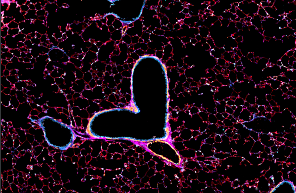
Multiplex spatial phenotyping enables to identify many different cell types simultaneously and map their organisation throughout the tumour tissue. Confocal microscopy then allowed them to examine the morphology and behaviour of endothelial cells in much greater detail.
By combining these complementary approaches, Petra and Pavel can investigate not only whether the therapy affects cancer cells, but also how it reshapes the surrounding tumour microenvironment.
The researchers chose the IMCF because it is the only academic provider of highly-multiplexed spatial proteomics services in Prague and offers expertise spanning sample preparation, imaging and image analysis.
"The spatial proteomics workflow was technically challenging," Petra explains. "The IMCF team helped us enormously with optimising the protocol and getting everything working."
The support extended well beyond image acquisition. The team helped establish an image analysis pipeline using QuPath and will continue supporting the researchers as they analyse their large and complex datasets.
"We've generated a huge amount of data," Petra says. "Collecting it is only the beginning. Now we need to extract the biological answers. We're also planning to share the data in a public repository so that other researchers may be able to answer different scientific questions using the same dataset."
Pavel particularly valued the opportunity to learn new skills and techniques throughout the project.
"It was my first time writing a grant application," he says. "Applying for canSERV was a great experience because I learned how a research project is run from the inside. I've also made valuable contacts and gained hands-on experience with multiplex and confocal imaging."
For Petra, one of the greatest benefits of the project has been establishing a long-term collaboration with the IMCF.
"Our institute doesn't have this type of microscopy infrastructure, so working closely with the IMCF has been incredibly valuable," she says. "The team has expertise across many different sample types and imaging approaches, and they've been very supportive throughout the project. We hope to continue collaborating with them in future studies."
Although the work is still ongoing, the researchers' first results are encouraging. By combining advanced imaging technologies with expert support, they are gaining a more comprehensive picture of how both cancer cells and the surrounding healthy tissue respond to treatment. Ultimately, they hope these insights will help explain why promising anti-cancer therapies sometimes lose their effectiveness—and contribute to the development of more durable treatments in the future.
"Applying for canSERV was a great experience because I learned how a research project is run from the inside. I've also made valuable contacts and gained hands-on experience with multiplex and confocal imaging."
-- Pavel Jakoubě, from the Institute of Biotechnology of the Czech Academy of Sciences
October 12, 2026
The second edition of the EVOLVE Mentoring Programme has come to a close, marking the end of several months of mentoring, knowledge exchange…
October 8, 2026
This September, Euro-BioImaging was part of the Data Stewardship goes Germany (DSgG) Community Meeting in Cologne. Under the theme “from going to…

October 7, 2026
On 28–29 September 2026, the growing French volume electron microscopy (vEM) community came together in Bordeaux for the first France VolumeEM Scientific Days.