
March 30, 2026
Euro-BioImaging at the EOSC ESFRI meeting in Milan
Euro-BioImaging was delighted to attend the ESFRI/EOSC Policy Workshop on “EOSC and Research Infrastructures: Opportunities and Strategies” in Milan from March 15-16, represented by…

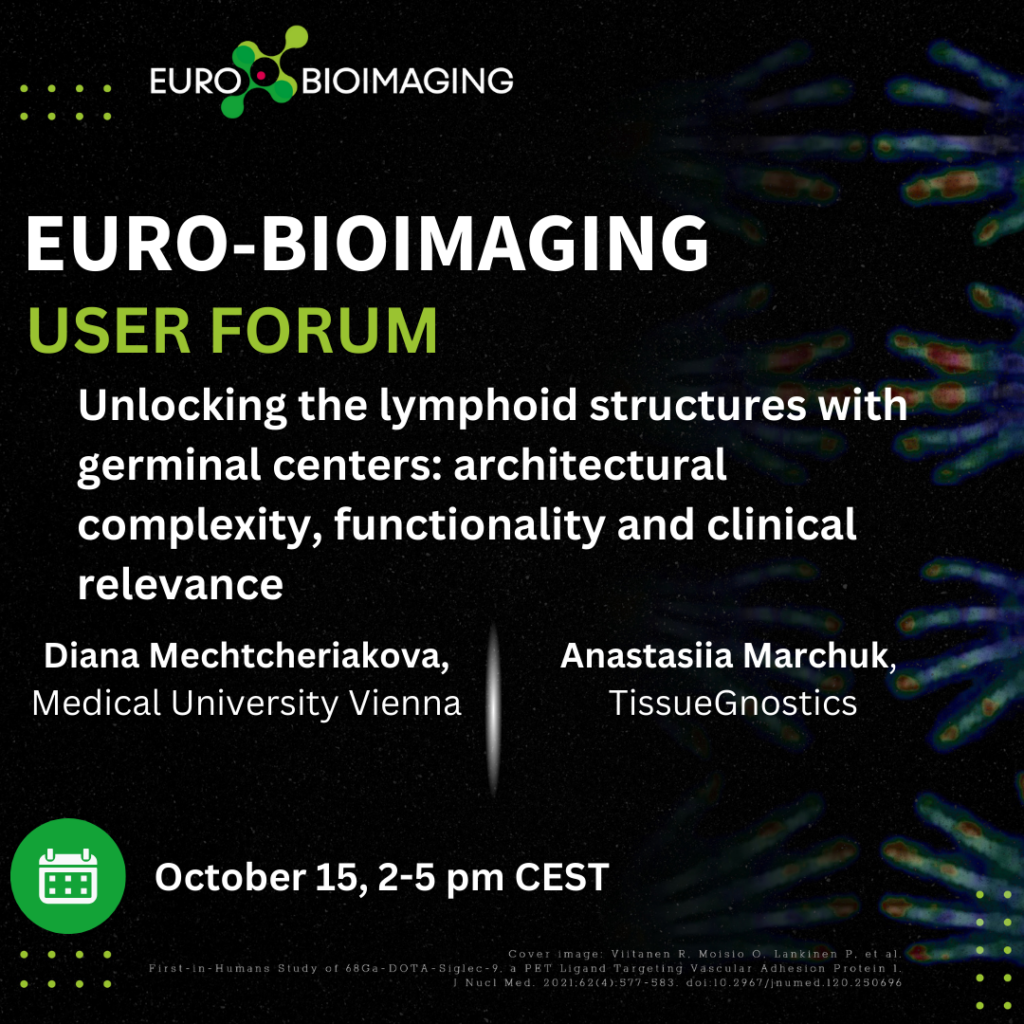
Imaging supports advances in Immunology and Inflammation research. The Euro-BioImaging User Forum “Focus on Immunology” will showcase cutting edge research in this domain from both a scientific and technical point of view. At this event, Diana Mechtcheriakova, Medical University Vienna, and Anastasiia Marchuk, TissueGnostics, share their collaboration to dissect lymphoid structures at tumor sites with AI-assisted image analysis and data mining, and Baubak Bajoghli will present the expertise of the Austrian BioImaging/CMI Node.
What: Euro-BioImaging User Forum “Focus on Immunology”
When: October 15, 2024, from 14:00-17:00 CEST
Where: Online
Unlocking the lymphoid structures with germinal centers: architectural complexity, functionality and clinical relevance
Diana Mechtcheriakova, Medical University Vienna
Anastasiia Marchuk, TissueGnostics
Baubak Bajoghli, Austria BioImaging Node
Lymphoid structures (LS) are integral part of the immune system and play an important role in the protection against pathogens as well as “foreign” objects such as cancer cells in terms of powerful humoral immune responses. The master regulator of LS with active germinal centers is the enzyme AID that drives the production of high affinity antibodies of various isotypes by plasma and memory B cells. LS are located not only in secondary lymphoid organs but are also formed in tissue sites with chronic inflammation and cancer. Understanding of the appearance, maintenance and functional activities of ectopic LS at the tumor site is of high relevance in translational research. Presence of LS at the tumor site was found to be associated with better clinical outcome in more than ten types of cancer. Important discoveries in respect of colorectal cancer with liver metastasis are based on our research.
Dissecting the complex immunological imprint of LS dictates the necessity for innovative methodology and state-of-the-art analytical solutions. Central to this is the quantitative analysis of LS using spatial tissue image cytometry (TissueFAXS platform), which allows to automatically scan the stained tissue sections and then, on the single-cell level, perform quantitative assessment of marker-positive cells within the native tissue environment. Staining-derived variables are used for alignment with clinicopathological parameters for patient stratification and prediction of disease outcome.
Technical aspects
Spatial tissue image cytometry permits to determine the in-situ phenotype of individual cells as well as histological entities. Using TissueFAXS microscopy platform, the time-consuming and potentially error-prone human evaluation of stained histological sections can be approached with an automated and reproducible technology. Image analysis software packages, such as Histo/TissueQuest and StrataQuest, allow to extract tissue-encrypted data by automated detection of single cells, marker quantification as well as identification of tissue structures. The measurements can be performed for more than 20 parameters per cell and marker and exported as numerical data. To further fuel data-rich translational research, new solutions are launched soon which feature AI-assisted image analysis and data mining.
March 30, 2026
Euro-BioImaging was delighted to attend the ESFRI/EOSC Policy Workshop on “EOSC and Research Infrastructures: Opportunities and Strategies” in Milan from March 15-16, represented by…
March 30, 2026
Euro-BioImaging was delighted to attend the Public Awareness & Engagement of Research Infrastructures (PAERI) conference, represented by External Communications Officer, Marianna Childress-Poli. This year’s…

March 26, 2026
The Madrid Advanced Microscopy Center (MAdMiC) is the first Euro-BioImaging Node in Madrid (Spain). It is formed through the collaboration and close work of…