The Danish BioImaging Infrastructure (DBI) contains imaging facilities across different Universities and Research Institutions in Denmark. During the European Microscopy Congress, members of the Euro-BioImaging and Global BioImaging team had a chance to visit the Core Facility for Integrated Microscopy (CFIM) at the University of Copenhagen and meet with DBI Director Clara Prats and DBI Coordinator Sonia Diaz Garcia. Here’s an article from Johanna Bischof to share this experience with you.
From light microscopy systems to laser systems
The CFIM is a large and well-organised facility, combining a range of light microscopy systems, from two advanced lightsheet systems, slide scanners, confocal, and super-resolution systems, alongside Electron Microscopy systems, including TEM, cryo-EM, and FIB-SEM systems. The users of the facility receive dedicated support from the technical experts in the facility.
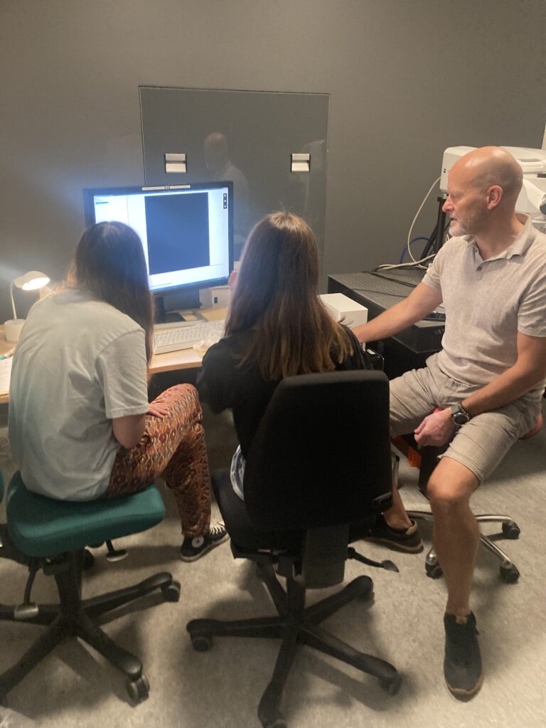
We could directly see the dedication of the team to quality maintenance of their systems, as Application Specialist Thomas Braunstein was training new facility staff on calibrating and measuring performance of one of the confocal systems during the visit.
Danish BioImaging Application Specialist Thomas Braunstein training new facility staff on how to calibrate the light microscopy machines.
Particularly impressive in the facility are a specialised equipment "corridor" housing all laser systems outside of the microscope rooms, into which the laser light is fed. This allows for better temperature control of the laser environment, improving lifetime, and leading to more efficient space use.
Managing user access
The facility also features a large screen displaying all systems bookings for the week right at the entrance of the facility. This allows the expert staff to plan their time and support all users who need their help in running the systems, and it contributes to monitoring of instrument usage.
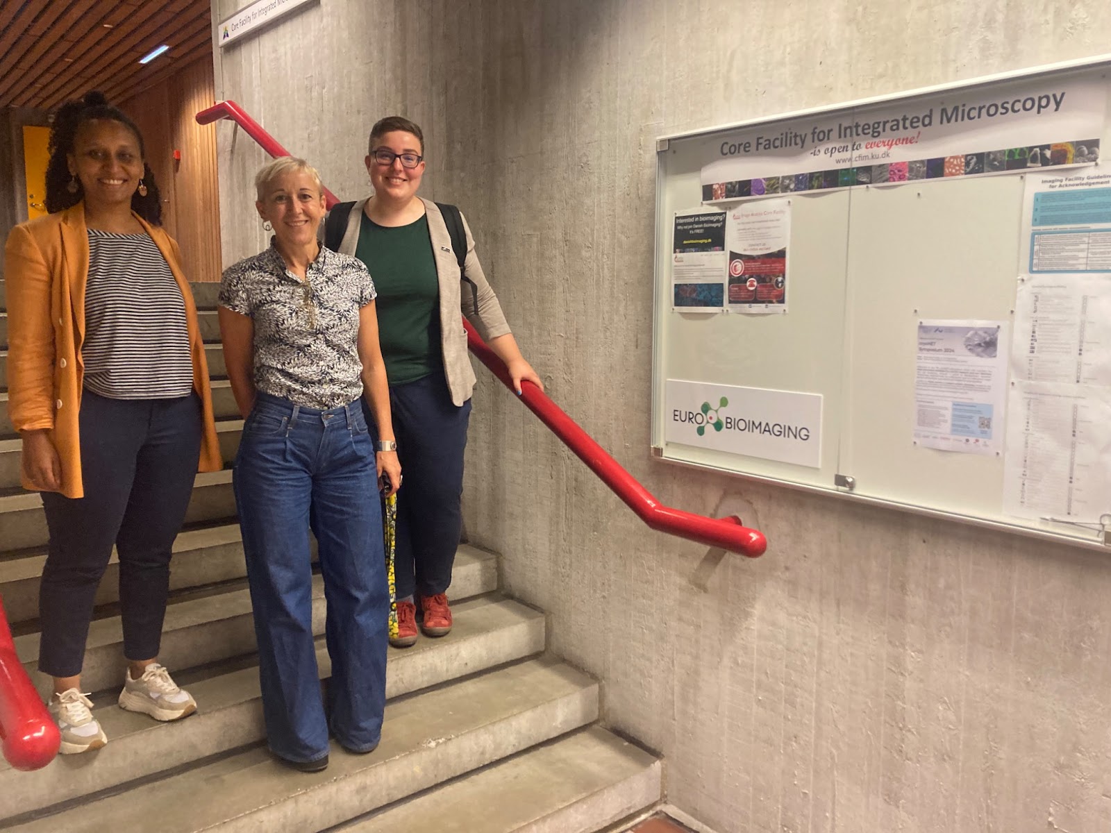
From left to right: Yara Reis, Global BioImaging, Clara Prats, Danish BioImaging, and Johanna Bischof, Euro-BioImaging, in front of the entrance to the CFIM facility. Photo by Sonia Diaz Garcia.
Image Data Analysis as a core service
The CFIM also offers open access image analysis services, and hosts the national DBI-INFRA Image Analysis Core Facility, a team of three image analysts/programmers implementing new image analysis tools and workflows to bioimaging scientists across the country and beyond.
Image analysis will also play an important role in the upcoming Molecules2Human bootcamp (M2H) - a two-week immersive training course combining biological and biomedical imaging. M2H is a special project, coordinated by Global BioImaging with funding from the Chan Zuckerberg Initiative.
Hosting a prestigious international microscopy “bootcamp”
Danish BioImaging was chosen as the host for the M2H bootcamp in a competitive selection among different Euro-BioImaging Nodes for its unique integrated and project-focussed training program designed for the bootcamp. M2H will take place in September and welcome 24 participants from around the globe for training in Aarhus and Copenhagen with local and global experts. It was great to get the latest update on the planning for the course from the organising team - M2H is certainly set up for success with an enthusiastic and expert training team.
Future perspectives
The visit also presented a chance to discuss future plans and sustainability considerations for Danish BioImaging, and to work to identify pathways for the Danish BioImaging community to get even more engaged in Euro-BioImaging.
Many thanks to Clara and Sonia for making the visit possible!
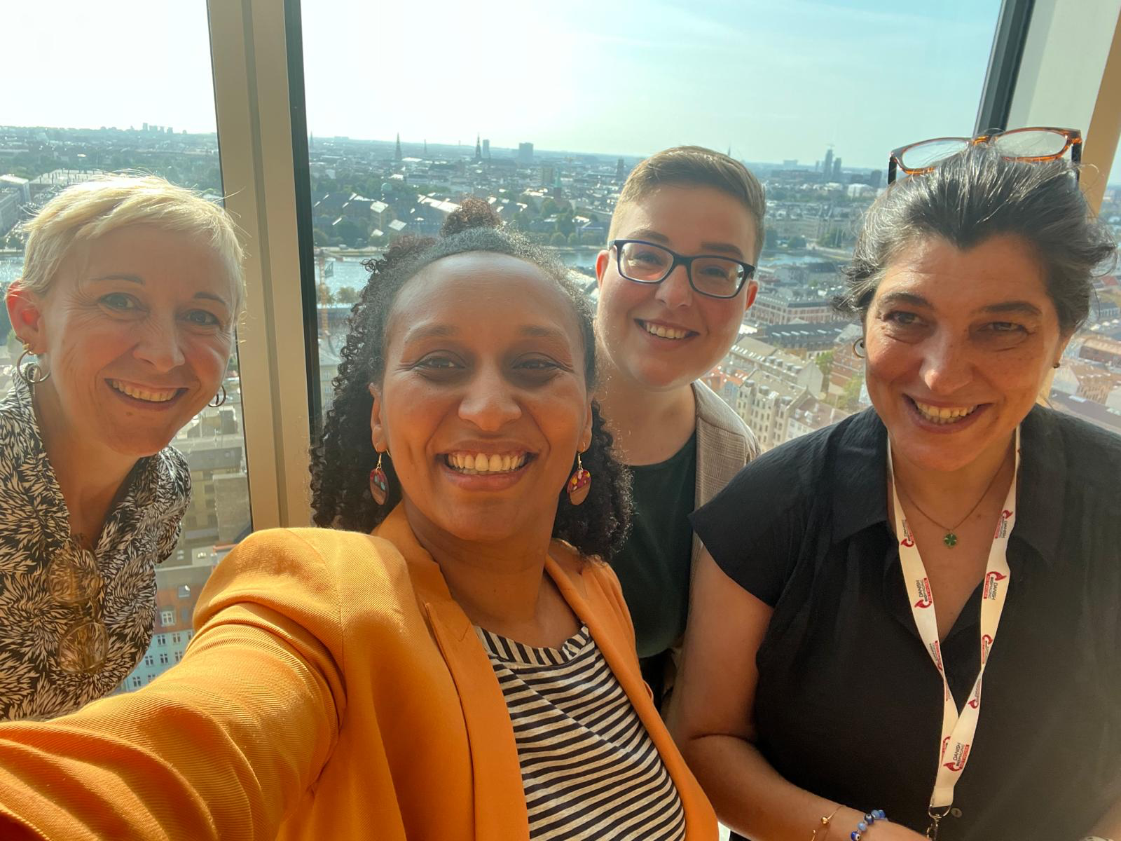
A visit to Danish BioImaging. Left to right: Clara Prats, Yara Reis, Johanna Bischof, Sonia Garcia
More news from Euro-BioImaging
July 21, 2026
Building Skills Across Europe: EVOLVE Supports the Second Edition of the Distributed Image Analysis Training Course
The second edition of the “Introduction to Image Analysis with Python for Life Scientists” course marked another successful milestone in the development of Euro-BioImaging’s distributed training model.
Strengthening research-industry partnerships: results from the EVOLVE industry job shadowing pilot
The EVOLVE project, supported by European Union funding, recently completed a pilot programme designed to deepen the relationships between academic research infrastructures…
Imaging-Driven Neuroscience, Responsible Research, and the Power of Advocacy: Highlights from FENS Forum 2026
Euro-BioImaging was pleased to participate in the FENS Forum 2026, Europe’s largest international gathering dedicated to advancing neuroscience (>8000 participants). Throughout…