The Madrid Advanced Microscopy Center (MAdMiC) is the first Euro-BioImaging Node in Madrid (Spain). It is formed through the collaboration and close work of four leading and well-established imaging facilities from two of Spain’s top research institutions: the Advanced Optical Microscopy and Preclinical Biomedicine Facilities at the Centro de Biología Molecular Severo Ochoa (CBM) and the Advanced Optical Microscopy and Cryo-Electron Microscopy Facilities at the Centro Nacional de Biotecnología (CNB).
MAdMiC operates as a fully integrated, co-located Node, staffed by 17 expert technicians, providing access to 26 microscopy systems and supporting over 25 microscopy techniques.
As of January 2026, the MAdMiC Node is ready to offer services to external users who apply for access to the facilities via the Euro-BioImaging web portal.
MAdMiC aims to specialise in cross-scale imaging, offering a comprehensive portfolio of techniques and expertise that enables researchers to address their biological questions across all relevant biological scales, with a particular focus on live samples and dynamic studies.
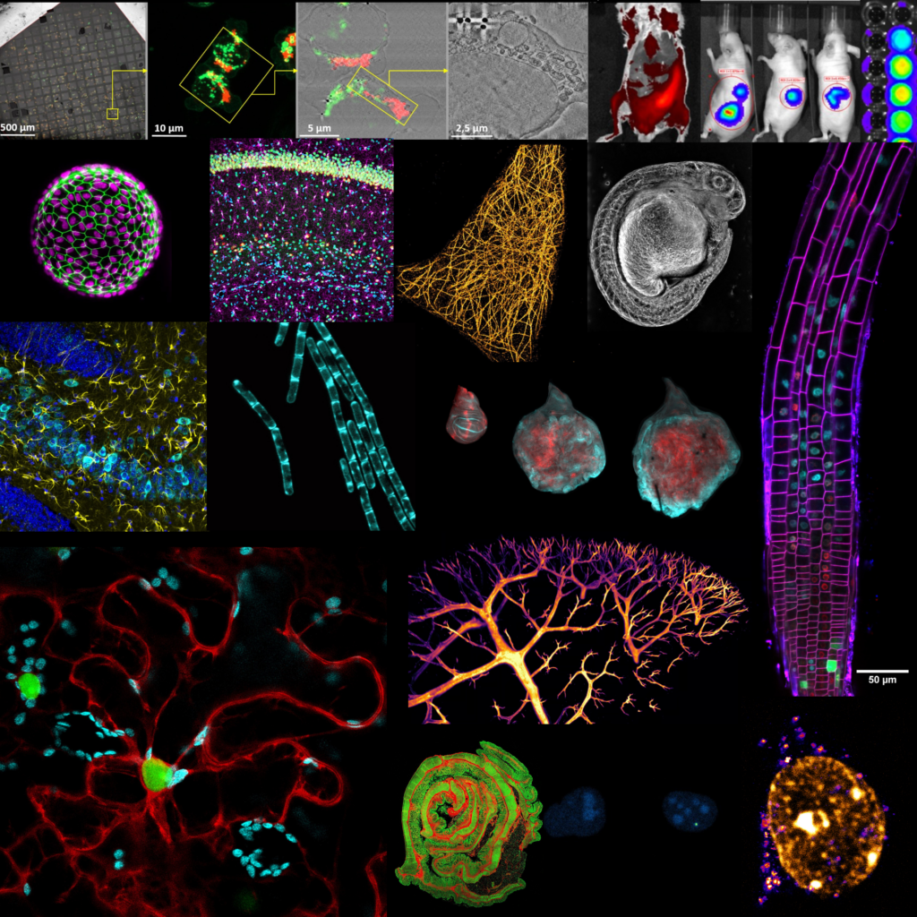
A collage of images showing the amazing range of imaging approaches available to users at the MAdMiC Node. Images courtesy of the MadMIC Node.
MAdMiC’s capabilities extend from vitrification-based cryo-electron microscopy (SEM, FIB and CLEM) to intravital and in vivo optical imaging, including advanced in vivo nanoscopy techniques (PALM, SoRa, TIRF), a broad range of functional microscopy approaches (FLIM, FRAP, FRET, FCS/FCCS), high-speed imaging, photomanipulation and life-oriented lightsheet microscopy.
What is their expertise?
”We are excited to offer Euro-BioImaging users our combined expertise. Our philosophy is to provide comprehensive, end-to-end support to researchers, accompanying them throughout the entire experimental workflow, from initial experimental design to data reporting and image publication. This includes advanced sample preparation, image acquisition and image pre-processing and analysis. In addition, we aim to cover multiple biological scales, enabling researchers to investigate their systems at different levels within the same Node and with the support of a single, integrated expert team,” explains Alvaro Sahun, Head of the CBM Advanced Optical Microscopy Facility.
“We offer advanced imaging approaches for a wide range of biological samples, including cultured cells, organoids, tissues, tissue explants, plants and bacteria,” affirms Ana Oña Blanco, Head of the CNB Advanced Optical Microscopy Facility. “Supported by a large multidisciplinary team of experts and a comprehensive portfolio of state-of-the-art imaging systems, we provide end-to-end support covering the full experimental workflow, from experimental design and advanced sample preparation to image acquisition, analysis and data visualization. Our cross-scale and life-imaging expertise, brought together within a single, co-located Euro-BioImaging Node, enables users to investigate biological processes across multiple spatial and temporal scales within one integrated infrastructure. Located in one of Southern Europe’s most dynamic innovation environments, the Node offers an optimal setting for cutting-edge biological imaging.”
Our philosophy is to provide comprehensive, end-to-end support to researchers, accompanying them throughout the entire experimental workflow, from initial experimental design to data reporting and image publication. This includes advanced sample preparation, image acquisition and image pre-processing and analysis. In addition, we aim to cover multiple biological scales, enabling researchers to investigate their systems at different levels within the same Node and with the support of a single, integrated expert team.”
-- Alvaro Sahun, Head of the CBM Advanced Optical Microcopy Facility
More news from Euro-BioImaging
March 26, 2026
Participate in the NFDI4BIOIMAGE Research Data Management Community Survey 2026
German BioImaging, within its work in the NFDI4BIOIMAGE consortium, and in collaboration with Euro-BioImaging ERIC, has launched a new survey to collect input about…
New EVOLVE Course: Communication and Management Skills for Imaging Core Facility Staff
Turin, Italy – 20–22 October 2026 Early career professionals working in imaging core facilities will soon have the opportunity to strengthen essential skills beyond…
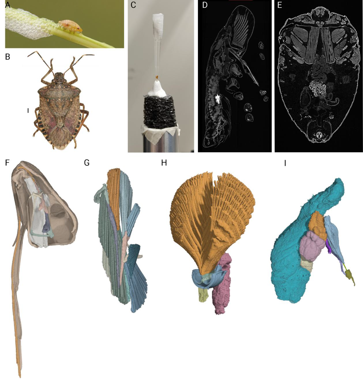
Use of synchrotron X-ray Phase-contrast MicroCT to investigate the reproductive system and the structures involved in foam-production in the spittlebug Philaenus spumarius
Use of synchrotron X-ray Phase-contrast MicroCT to investigate the reproductive system and the structures involved in foam-production in the spittlebug Philaenus spumarius The Olive…