
July 9, 2026
Euro-BioImaging Visits the NORMOLIM Node at the 2026 180°N Conference
In April 2026, the Med-Hub Head of Operations Alessandra Viale attended the 2026 180°N Conference, held at the beautiful Nye Hjorten Teater…

Are you interested in looking at tissues or other thick samples in high resolution? We spoke to Marc Tramier, a group leader at the Institute of Genetics & Development of the University of Rennes/INSERM/CNRS, and scientific director of MRic (Microscopy Rennes Imaging Centre), to learn more about Random Illumination Microscopy (RIM), a fast and easy to use microscopy technique with low phototoxicity. His facility, which is part of the Bretagne-Loire Node of France BioImaging , offers RIM as a Euro-BioImaging Proof-of-Concept study, and is now accepting applications for projects. Read more about this technology in the interview below.
We are today talking about Random Illumination Microscopy imaging. Please provide a short summary of this type of imaging and list some applications:
Marc Tramier: This microscopy method is a transfer from my colleague Thomas Mangeat (CBI Toulouse, France). The basic idea of Random Illumination Microscopy is to use the speckle of the illumination laser in wide field to create a structured illumination pattern at the diffraction limit. By varying the pattern from image to image using a diffracting element (in our case a SLM), we are able to acquire a stack of images (around 100 images) on a camera which corresponds to a cumulative homogeneous illumination. By resolving the inverse problem, we are able to reconstruct a super-resolved image at the focal plane with unprecedented optical sectioning. In comparison to conventional SIM, RIM is able to work in depth inside diffusive samples as the speckle is insensitive to diffusion.
The main application is to acquire super-resolved images in depth with very low amount of light at high speed. This makes a very nice compromise of z-sectioning and super-resolution with wide field illumination particularly adapted to thick live samples.
Tell us a bit more about a specific project that was done in your facility using this technology? What scientific questions were you addressing?
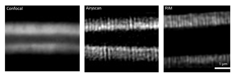
Marc Tramier: The method was first implemented by Thomas Mangeat and collaborators in Toulouse (Mangeat et al., Cell Rep Methods. 2021 Apr 30;1(1):100009. doi: 10.1016/j.crmeth.2021.100009 ). In our facility, after the transfer of the prototype to offer it as a user-friendly system, we are able to image microvilli of intestine in c-elegans (depth > 50µm) having a spatial resolution of around 100 nm. This structure is impossible to be revealed by conventional confocal microscopy. Before the use of RIM, only airyscan approach allowed us to resolve the microvilli but with higher illumination power (photobleaching of the sample) and longer acquisition time (around 10 times more). Now with RIM, we are able to follow microvilli in the living C–elegans at the second time-scale during several minutes.
What are the advantages of RIM?
Marc Tramier: To our knowledge, RIM is one of the powerful method to achieve the best resolution at high speed with very low phototoxicity.
Another advantage of RIM is that it is not a difficult technique. For a trained microscopist, it’s very easy. The main difficulty would be to find the parameters for the reconstruction, there may be some difficulties in data analysis. At our facility we have a user-friendly prototype of this system and it’s available in open access through Euro-BioImaging. Similar systems are also available in Toulouse and Marseille.
What other services do you provide in your facility that would be useful in combination with this type of imaging?
Marc: We have a complete set of microscopy methods for live sample investigation, from wide-field to light sheet including spinning disk, confocal and airyscan.
How to apply to use RIM:
Random Illumination Microscopy is part of the Euro-BioImaging Proof-of-Concept study, in collaboration with our Nodes. The Proof-of-Concept study makes it possible to introduce exciting, new imaging technologies to our portfolio that were previously unavailable via our network. We are currently accepting applications to use these technologies as part of the Proof-of-Concept study. Be part of this study - and contribute to community-wide continuous technological innovation!
All scientists, regardless of their affiliation, area of expertise or field of activity can benefit from Euro-BioImaging’s pan-European open access services. Potential users of these new technologies are encouraged to submit project proposals via our website. To do so, you can log in to access our application platform, choose the technology you want to use and the facility you wish to visit, then submit your proposal. All applications will be processed by the Euro-BioImaging Hub. As usual, users will benefit from advice and guidance by technical experts working at the Nodes, training opportunities, and data management services.
For more information: info@eurobioimaging.eu
July 9, 2026
In April 2026, the Med-Hub Head of Operations Alessandra Viale attended the 2026 180°N Conference, held at the beautiful Nye Hjorten Teater…
July 7, 2026
Armed conflicts generate long-lasting environmental contamination that extends well beyond the duration of military operations. The release of heavy metals such as Arsenic,…
July 6, 2026
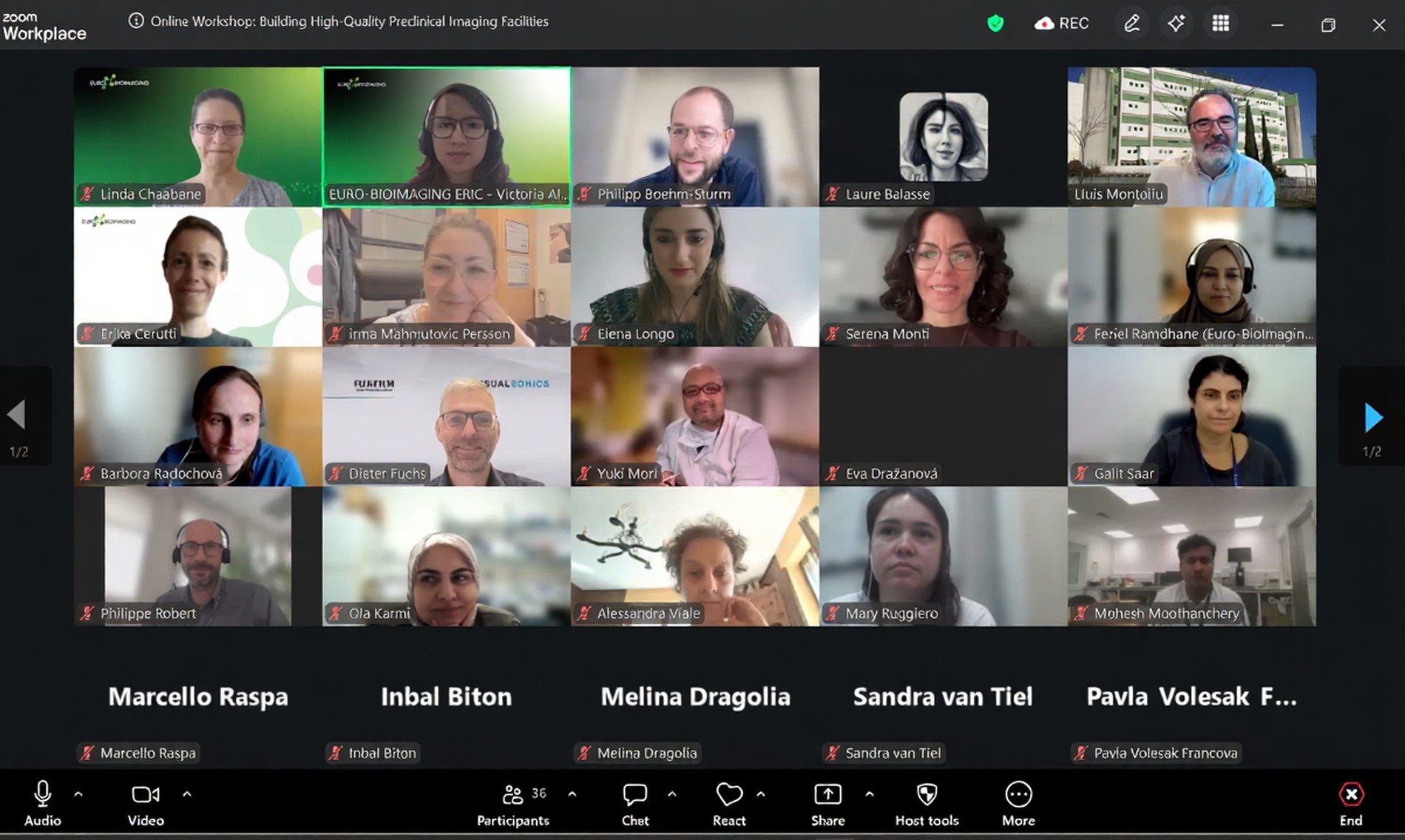
On 2 July 2026, Euro-BioImaging hosted the online EVOLVE workshop “Building High-Quality Preclinical Imaging Facilities”, bringing together approximately 40 imaging facility staff and…