
October 16, 2026
RAISE Hub: One gateway to Europe’s AI in Science Community
A new online platform to connect researchers, infrastructures, and funders to accelerate AI scientific discovery across Europe Dublin, Ireland, 15 October…



Developing new experimental models for cancer research requires not only innovative technologies, but also the ability to see what is happening inside complex biological systems. Through canSERV, Bulgarian SME MatriChem accessed advanced light and electron microscopy expertise at the Imaging Methods Core Facility (IMCF) at BIOCEV, part of the Advanced Light & Electron Microscopy Prague Node of Euro-BioImaging, using complementary imaging approaches to investigate a new 3D-bioprinted model of the tumour microenvironment and identify important next steps in its development.
Understanding how cancer cells interact with their surrounding environment is important for studying cancer progression and developing new therapeutic strategies. Yet these complex interactions are difficult to reproduce in conventional two-dimensional cell culture models.
MatriChem Ltd, a Bulgarian SME specialising in biomaterials and bioprinting, is working with researchers at the Medical University of Plovdiv to develop a three-dimensional bioprinted model that recreates key features of the interface between a tumour and blood vessels.
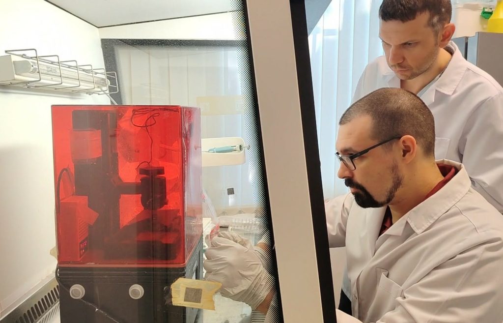
The 3DCAM platform combines cancer cells, endothelial cells and biomaterials in a structured 3D environment. In the longer term, the team aims to establish a robust experimental platform that can be used to investigate processes associated with tumour vascularisation and metastasis, and potentially support preclinical studies.
At this early stage of development, however, understanding the structure of the model and the interactions between cells and the surrounding biomaterial was an important challenge. This required imaging capabilities and expertise beyond those readily available to the company.
Through the canSERV project, MatriChem received access to the IMCF at BIOCEV, part of the Euro-BioImaging Advanced Light and Electron Microscopy Node Prague CZ.
The project brought together the company's expertise in bioprinting and biomaterials with the facility's experience in microscopy imaging and ultrastructural analysis of bio-composite samples. With the model combining cancer cell biology and mechanobiology with bioengineering and manufacturing technology, detailed planning between the two teams was an important part of the collaboration.
Anticipating the challenges presented by these complex samples, the teams worked together to optimise sample preparation, experimental conditions and the logistics of sample delivery. In particular, they needed to find a compromise between conditions suitable for manufacturing the samples, maintaining cell viability and growth, and preparing them for microscopy. This careful planning allowed the project to generate useful results from the available samples without requiring repeated sample production – an important consideration in terms of both time and cost.
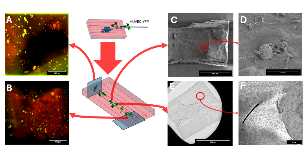
The team applied multiple complementary imaging approaches, ranging from stereomicroscopy and 3D fluorescence confocal microscopy to scanning and transmission electron microscopy.
Confocal microscopy was initially used to examine the distribution of cells within the bioprinted structures. The work also demonstrated some of the challenges inherent in developing and characterising a new biological model. Fluorescence from one of the labelled cell populations was not sufficiently clear to distinguish the different cell types as originally planned. Rather than providing the complete functional characterisation initially envisaged, this first imaging step therefore highlighted an important issue for further optimisation.
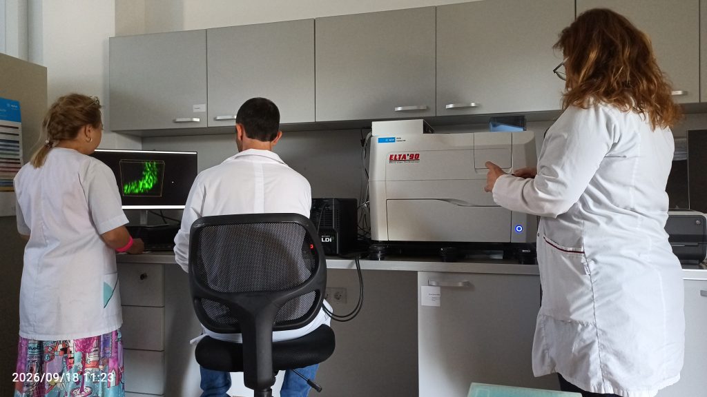
Scanning and transmission electron microscopy subsequently allowed the samples to be investigated at much higher resolution. These complementary approaches provided detailed information about the structure and porosity of different hydrogels, the organisation of cells within the model, and the interfaces and contacts between cells and the surrounding material.
“For a small company like MatriChem, the biggest value of accessing advanced microscopy infrastructure was being able to make development decisions based on much deeper evidence than we could generate on our own. At this stage, knowing where not to invest time and resources is just as important as knowing what to pursue. When those decisions are informed by results obtained using well-established characterisation protocols, we can move forward with much greater confidence in the next steps of our product development.”
– Murad Redzheb, Founder & Managing Director, MatriChem
For an experimental platform at this stage of development, identifying limitations can be as valuable as confirming what already works.
The microscopy results helped the team identify several parameters requiring further optimisation, including cell viability and density within the hydrogel, cell attachment and growth, and fluorescence labelling. The joint work with the IMCF at BIOCEV also helped the MatriChem team develop its understanding of the challenges involved in confocal imaging of thick, cell-laden hydrogels, better equipping them to characterise biofabricated constructs in the future.
At the same time, electron microscopy provided valuable insight into hydrogel structure and cell–material interactions. Bringing the results from the different imaging modalities together was instrumental in identifying key factors for further optimisation and functional development of the model.
The learning went both ways. For the IMCF team, working with these unusual samples provided valuable experience with the specific limitations of the different imaging methods, troubleshooting and optimisation of imaging workflows, and interpreting results consolidated across different microscopy modalities.
“Combining complementary imaging approaches was instrumental in characterising this challenging in vitro cancer model and identifying key factors for its further optimisation. The interdisciplinary collaboration was also a valuable exchange of experience for both teams.”
– David Liebl, Electron Microscopy Specialist, Imaging Methods Core Facility at BIOCEV
Together, the results provide a basis for refining both the 3DCAM model and the approaches used to characterise it. The work represents an important first step towards establishing a more robust pipeline for producing and analysing the 3D-bioprinted system.
The collaboration has also resulted in a joint scientific publication in Frontiers in Bioengineering and Biotechnology, bringing together researchers from MatriChem and the Plovdiv Medical University in Bulgaria with microscopy experts from the IMCF at BIOCEV in Prague. The publication provides a scientific account of the development and characterisation of the 3DCAM model and the results of the collaborative work.
For small companies developing new technologies, gaining access to specialised instrumentation is only part of the challenge. Equally important is access to scientists with the expertise to adapt imaging strategies to unusual samples, troubleshoot unexpected results and determine which approaches can provide useful information.
By providing transnational access to the microscopy expertise and technologies available at the Prague facility, canSERV allowed MatriChem to test and refine approaches for characterising its new model without first having to establish these highly specialised capabilities in-house.
The project illustrates how access to European research infrastructures can help SMEs explore ambitious new approaches at an early stage of development. In this case, the value was not simply in producing images, but in reducing uncertainty: identifying technical challenges, gaining new information about the experimental system and providing a clearer basis for the next development steps.
The resulting joint publication is another important outcome of this type of access: connecting complementary expertise across a Bulgarian SME, academic researchers and a European microscopy facility, and turning an exploratory access project into jointly generated scientific and technical knowledge.
Through canSERV - an EU-funded INFRA-SERV project (Grant number 101058620) - researchers from academia and industry can access cutting-edge cancer research services across Europe, supporting projects ranging from fundamental cancer biology to the development of new technologies and therapeutic approaches.
October 16, 2026
A new online platform to connect researchers, infrastructures, and funders to accelerate AI scientific discovery across Europe Dublin, Ireland, 15 October…
October 16, 2026
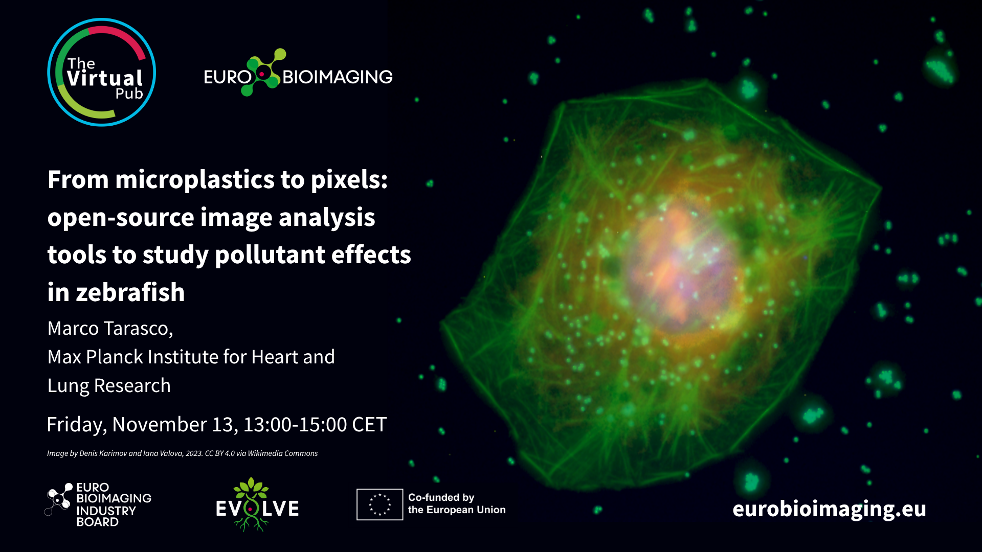
At the Special Edition Virtual Pub, “Imaging Micro- and Nanoplastics,” Euro-BioImaging users, Node experts and Industry partners will explore how advanced microscopy, clinical…
October 16, 2026
At the Special Edition Virtual Pub, “Imaging Micro- and Nanoplastics,” Euro-BioImaging users, Node experts and Industry partners will explore how advanced microscopy, clinical…