
October 16, 2026
RAISE Hub: One gateway to Europe’s AI in Science Community
A new online platform to connect researchers, infrastructures, and funders to accelerate AI scientific discovery across Europe Dublin, Ireland, 15 October…



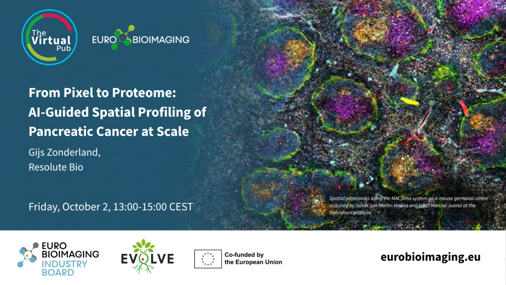
Spatial proteomics is rapidly transforming the way researchers study complex biological systems. To explore the latest developments in this fast-moving field, Euro-BioImaging is organising a Special Edition Virtual Pub dedicated to Spatial Proteomics on Friday, October 2, from 13:00–15:00 CEST, bringing together experts from academia and industry.
At this event, Gijs Zonderland, Resolute Bio, will explain how AI-guided image analysis contributed to a spatial proteomic atlas spanning PDAC progression from the earliest molecular transitions in precursor lesions to advanced metastatic disease, identifying potential biomarkers and actionable therapeutic targets along the way.
From Pixel to Proteome: AI-Guided Spatial Profiling of Pancreatic Cancer at Scale
By: Gijs Zonderland, Resolute Bio
Resolving tissue architecture at molecular resolution is invaluable for translational research. Pathology foundation models have revolutionized H&E image analysis, but they remain limited to the morphological information encoded in the pixel. Connecting these visual representations to the underlying molecular phenotype requires spatially matched, high-throughput experimental ground truth. Here, we close this gap by coupling AI-guided image analysis with spatially resolved, unbiased protein quantification by mass spectrometry (MS). We applied this platform in pancreatic ductal adenocarcinoma (PDAC) to map molecular events across the full disease trajectory, from non-malignant precursor lesions to metastatic disease. Using computational pathology driven by foundation models, we located pancreatic intraepithelial neoplasia (PanIN), the most common precursor lesion, alongside matched histologically normal ductal epithelium. Based on these biological regions, we performed Deep Visual Proteomics (DVP) to isolate ~100 epithelial cells using targeted laser microdissection followed by high-resolution MS. Across 9,181 quantified proteins, we showed that proteome-level reprogramming precedes histological transformation. To enable high-throughput spatial profiling, we further scaled the platform to match proteomes directly to the image units of computational pathology (tileDVP). Applying this approach to a cohort of matched primary and metastatic PDAC samples, we profiled ~15–40 cells per region across 55 FFPE blocks from 10 patients, generating a total of 2,542 proteomic measurements. The resulting spatial proteomic atlas spans PDAC progression from the earliest molecular transitions in precursor lesions to advanced metastatic disease and identifies potential biomarkers and actionable therapeutic targets along the way.
October 16, 2026
A new online platform to connect researchers, infrastructures, and funders to accelerate AI scientific discovery across Europe Dublin, Ireland, 15 October…
October 16, 2026
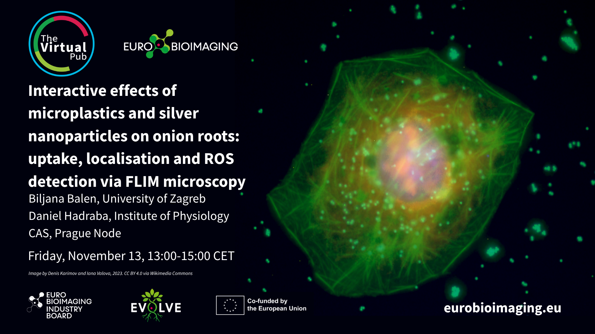
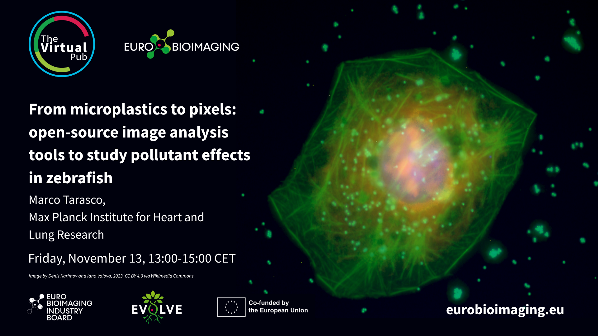
At the Special Edition Virtual Pub, “Imaging Micro- and Nanoplastics,” Euro-BioImaging users, Node experts and Industry partners will explore how advanced microscopy, clinical…
October 16, 2026
At the Special Edition Virtual Pub, “Imaging Micro- and Nanoplastics,” Euro-BioImaging users, Node experts and Industry partners will explore how advanced microscopy, clinical…