
July 9, 2026
Euro-BioImaging Visits the NORMOLIM Node at the 2026 180°N Conference
In April 2026, the Med-Hub Head of Operations Alessandra Viale attended the 2026 180°N Conference, held at the beautiful Nye Hjorten Teater…


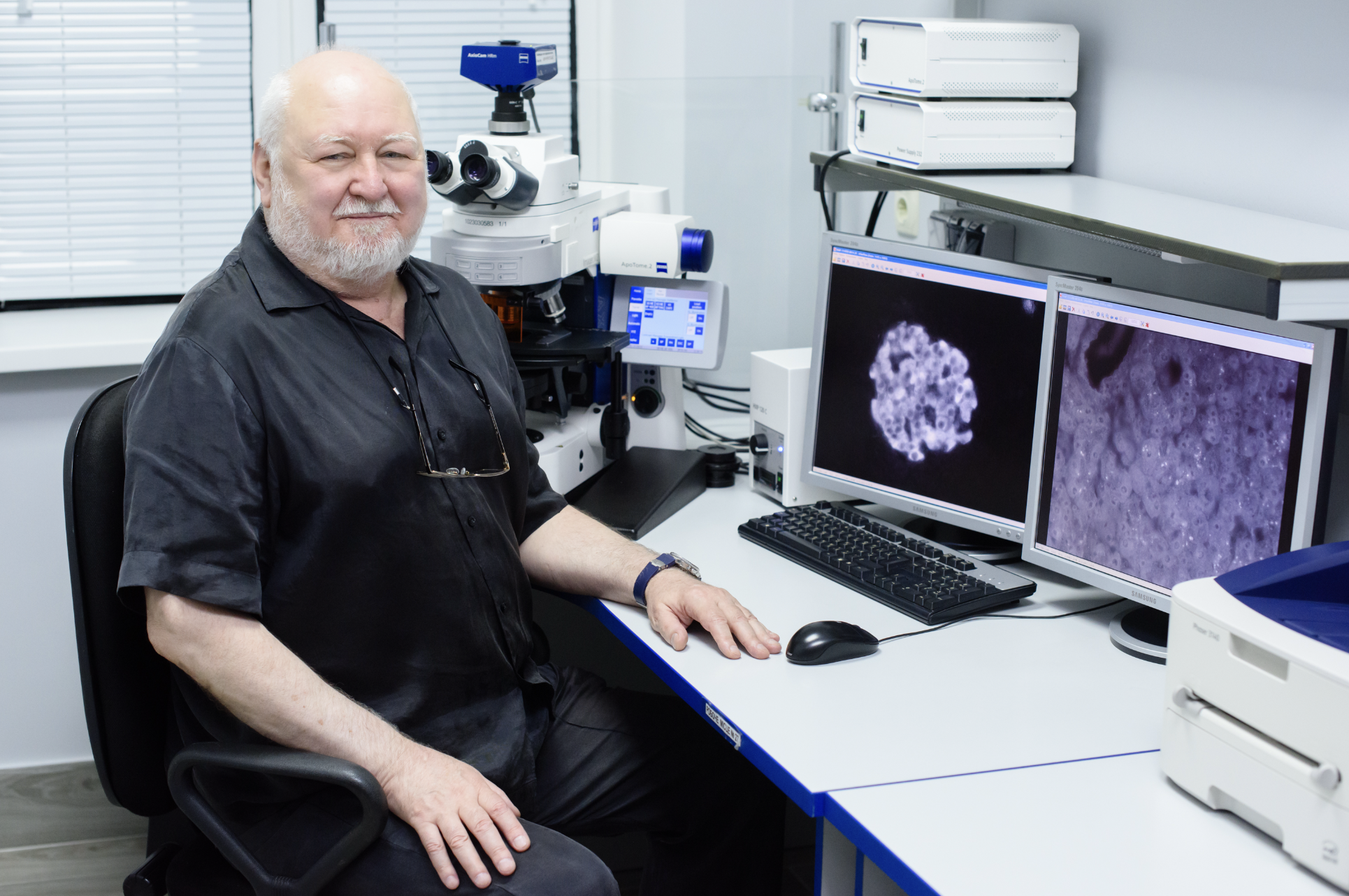
My name is Camilo Guzmán and I am the manager of the Finnish Advanced Light Microscopy Node of Euro-BioImaging. I have a background both in physics and in biology and I have been working on microscopy topics for over 17 years now. Our node has imaging facilities in Helsinki, Oulu and Turku and I am based in Turku where I take care of administrative tasks, while also providing support to different users on super-resolution microscopy, fluorescence dynamics and several biophysics methods.
Atomic force microscope (AFM) is a type of scanning probe microscopy technique capable of imaging surfaces down to a sub-nanometer resolution. This technique works by “touching” the surface with a mechanical probe that “feels” the changes in topography as the system scans the sample thanks to piezoelectric materials that allow for high precision movements. Besides imaging the topography of all kinds of materials, AFM is also used to test the mechanical properties of the sample in a process called force spectroscopy. With this process, it is possible for instance to test binding forces between proteins or cells, or to perform indentation on a sample and obtain its Young's modulus as a measure of the sample stiffness. In biology, AFM can be performed thanks to the introduction of a liquid cell that allows working on many samples that require a liquid media and even on living cells. Some of the latest developments of AFM allow very high speed of imaging while still keeping a nanometer resolution, this has been used to study for example the movement of kinesins along microtubules.

A close-up of the Atomic Force Microscopy (AFM) machine at the Finnish Advanced Light Microscopy Node

In our facility AFM has been used to test the mechanical properties of different materials such as hydrogels used in cell culture [1], different kinds of mammalian tissue [2], and even individual cells. In all these cases AFM has been essential to obtain the Young’s modulus of the sample and in such way describe its stiffness as a function of location, treatment or present mutations. We have also used AFM to image for instance nanoparticles and erythrocytes and to measure adhesion of cells to substrates.
For experiments where molecular binding forces need to be measured or where the mechanical properties of a material need to be characterised at a micrometric scale and under very low forces (pN to nN), AFM is one of the most suitable instruments to use. AFM not only is very precise, but it is highly versatile to different materials and samples and furthermore it can be combined with fluorescence microscopy to give a full description of the sample that is being tested.
AFM is a type of microscopy that requires a lot of patience and time, the probes used to image or mechanically test your sample are extremely fragile and need to be handled carefully. Furthermore, the piezoelectric materials used for scanning are also very fragile and therefore the user needs to know very well what he/she is doing to avoid crashing the system and breaking the scanner. Finally, alignment and calibration are time consuming activities that are critical to the success of the experiment and therefore need to be performed carefully. Training users on this technique requires a lot of time but once the users are capable of working independently, the technique is highly rewarding, and they will soon be obtaining very interesting results.
In our facility AFM can be used in combination with confocal microscopy as the two systems are merged and can be used one after the other or even at the same time. We provide training to our users in all the steps of the experiment and we advise them on the most suitable probes for the needs of their specific experiments. Also, all the other light microscopy techniques we offer are of course available on separate instruments.
Users should come to our facility not only because we have many years of experience using AFM in all its modalities and with all sorts of samples but also because we provide a fully personalized service where we will make sure that the user really knows what he/she is doing and what is the best approach to solve his/her scientific question. Also, it is quite unique that in our facility AFM and confocal fluorescence microscopy can be combined to obtain much more information about the same sample. Another advantage on our local system is that it is equipped with a CellHesion module that allows 100 um of movement in the Z range, enabling for instance adhesion measurements with an entire cell attached to the cantilever.
References:
[1] Barber-Pérez, N. et al. Mechano-responsiveness of fibrillar adhesions on stiffness-gradient gels. J. Cell. Sci. 133, (2020).
[2] Peuhu, E. et al. SHARPIN regulates collagen architecture and ductal outgrowth in the developing mouse mammary gland. EMBO J. e201694387–18 (2016). doi:10.15252/embj.201694387
In July 2020, Euro-BioImaging launched a Proof-of-Concept study - in collaboration with its Nodes - making it possible to access six previously unavailable imaging technologies. This article – the fourth in a series - features interviews with Euro-BioImaging Node staff, shedding light on some potential applications of these technologies - and providing advice on how to get the most out of them. We are currently accepting applications to use these technologies as part of the Proof-of-Concept study.
All scientists, regardless of their affiliation, area of expertise or field of activity can benefit from Euro-BioImaging’s pan-European open access services.Potential users of these new technologies are encouraged to submit project proposals via our website. To do so, you can Login to access our application platform, choose the technology you want to use and the facility you wish to visit, then submit your proposal. All applications will be processed by the Euro-BioImaging Hub. As usual, users will benefit from advice and guidance by technical experts working at the Nodes, training opportunities, and data management services.
For more information: info@eurobioimaging.eu
July 9, 2026
In April 2026, the Med-Hub Head of Operations Alessandra Viale attended the 2026 180°N Conference, held at the beautiful Nye Hjorten Teater…
July 7, 2026
Armed conflicts generate long-lasting environmental contamination that extends well beyond the duration of military operations. The release of heavy metals such as Arsenic,…
July 6, 2026
On 2 July 2026, Euro-BioImaging hosted the online EVOLVE workshop “Building High-Quality Preclinical Imaging Facilities”, bringing together approximately 40 imaging facility staff and…