We are delighted to announce the winners of the “Winter” round of our Four Seasons of the Invisible Imaging Contest. Launched in early 2025, the competition highlights seasonally inspired imaging work that reveals the unseen beauty of the microscopic world. For “Winter,” we were treated to images reflecting the stillness and structure of the colder months, from frozen biological forms and delicate crystalline-like particles to scenes evoking warmth and togetherness in the microscopic realm.
We received a high number of creative entries, and our panel of six judges had the difficult task of selecting the winners based on both artistic quality and relevance to the seasonal theme. The competition was very tight, even resulting in a tie for second place among the top entries.
And the winners are...
The first prize goes to Claudia Victoria Benke of Phase Contrast Imaging Flagship Node Trieste for her image “Winter's Anatomy: When Lungs Become Trees”. The runners-up are Hélène Roberge of VIB-BIC Leuven for her image “Nano Ice Skaters”, and Karan Sharma of Stockholm University for her image “Liquid-liquid phase separation”.
Claudia will receive reimbursement of up to €1,000 to attend the scientific conference of her choice, and each runner-up will receive reimbursement of up to €500 to attend a scientific conference.
Check out the breathtaking winning images below, and stay tuned for upcoming opportunities to take part in future Euro-BioImaging imaging contests!
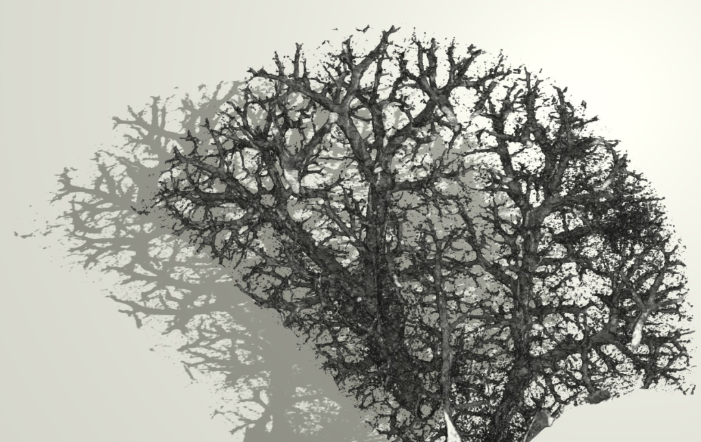
Winning image titled Winter’s Anatomy: When Lungs Become Trees, by Claudia Victoria Benke
Winter’s Anatomy: When Lungs Become Trees, by Claudia Victoria Benke, Phase Contrast Imaging Flagship Node, Trieste, Italy
Resembling a leafless tree in winter, this image reveals the branching airway system of a human lung. Captured post-mortem, the intricate network of bronchi and bronchioles appears suspended in stillness, its structure preserved while its function has ceased. Synchrotron phase-contrast computed tomography uncovers fine peripheral airways not visible with conventional imaging, exposing the lung’s hidden architecture in striking detail.
It is a propagation-based synchrotron phase-contrast computed tomography (PBI-CT) image acquired at the SYRMEP beamline of Elettra Synchrotron, followed by phase retrieval and 3D reconstruction.
The author thanks collaborators Christian Dullin, Davide Radaelli, Tommaso Bruscagin, Deborah Bonazza, Elena Longo, Philipp Nolte, Sam Bayat, Giuliana Tromba, and Stefano D’Errico, and acknowledges Euro-BioImaging ERIC for access to advanced imaging technologies through the Phase Contrast Flagship Node in Trieste, Italy.
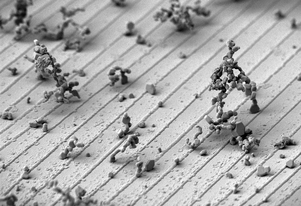
Runner-up image titled Nano Ice Skaters by Hélène Roberge
Nano Ice Skaters, by Hélène Roberge, VIB-BIC Leuven, Belgium
These frozen dust particles, imaged at the edge of a sample carrier during a cryo-SEM session, resemble tiny figures in motion, perhaps ice skating or engaged in a playful snowball fight. What might otherwise go unnoticed becomes a lively winter scene, where chance and imagination bring character to the microscopic world.
It is a cryo-scanning electron microscopy (cryo-SEM) image of a sample holder covered with frozen dust.
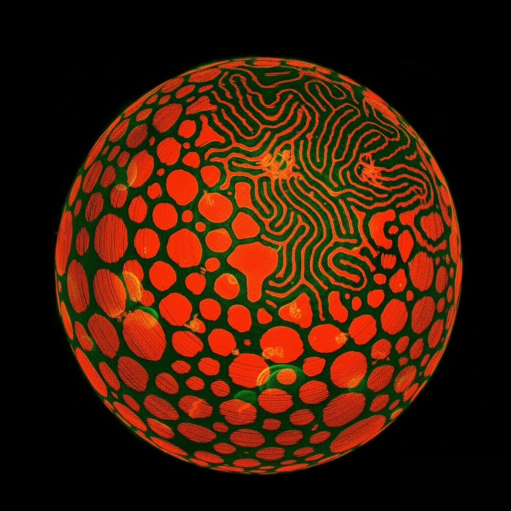
Runner-up image titled Liquid–Liquid Phase Separation by Karan Sharma
Liquid–Liquid Phase Separation, by Karan Sharma, Stockholm University, Sweden
The winter season and Christmas are closely linked, reflected here in a 3D projection of a confocal fluorescence microscopy image of a liquid–liquid phase-separated liposome. Red domains float within a green background, evoking familiar holiday colors. More than just phase separation, it feels like people coming together during the holidays, leaving behind daily rush and finding calm, warmth, and connection.
It is a projection of a 3D confocal fluorescence microscopy stack, illustrating two dyes that partition into separate liquid phases. The author acknowledges Fred Heberle and Milka Doktorova.
Congratulations to the winners!
Congratulations to the winners, and thank you to everyone who participated in the Winter round of Four Seasons of the Invisible! We will be sharing more stunning entries via our social media channels soon.
More news from Euro-BioImaging
July 24, 2026
A Multiplexed Vision: Highlights from the Special Edition Virtual Pub on Spatial Transcriptomics
On June 12th, Euro-BioImaging hosted its latest Special Edition Virtual Pub, bringing together over 200 researchers and technology developers working at the cutting…
Cellular Imaging Hungary Node expands expertise in advanced neurophotonics
The Euro-BioImaging Cellular Imaging Hungary Node has expanded its service portfolio with the addition of the BrainVisionCenter (BVC) in Budapest. Following a successful…
The UK Euro-BioImaging Node expands from seven to thirteen sites!
We’re delighted to announce that the UK Euro-BioImaging Node is expanding, growing from seven to thirteen sites following a successful upgrade application and…