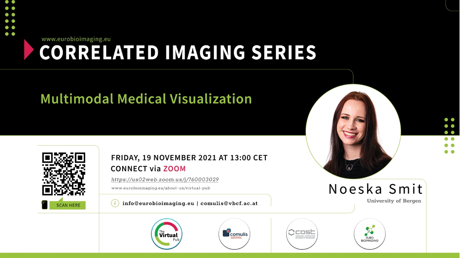
Correlated Imaging Series: Multimodal Medical Visualization
Published November 9, 2021

On Friday, November 19th at 13:00 CET, Noeska Smit of the University of Bergen delivers a lecture on how best to perform visual analysis of different imaging modalities, as part of the Correlated Imaging Series, brought to you by COMULIS and Euro-BioImaging.
Advances in medical imaging techniques are bringing more and more different imaging modalities that provide additional information on anatomy and physiology. For instance, a single patient can have a CT scan, PET scan, as well as an MRI scan with different weighted images. When there is more than one modality acquired, mental integration of the different contrasts between the different images becomes more challenging. Noeska Smit and her research team aspire to develop novel interactive visualization approaches for improved exploration, analysis, and communication of such multimodal medical imaging data. Their current focus in this context is on multi-parametric MR acquisitions. In this talk, Noeska Smit will provide a general introduction to medical visualization and highlight several visualization applications aimed at improved visual analysis for multimodal imaging data related to ongoing gynecological cancer and multiple sclerosis research.
About Noeska Smit:
Noeska Smit is a tenure-track Associate Professor in the visualization research group at the University of Bergen, Norway, since 2017, where she leads a team researching multimodal medical visualization. She is also affiliated with the Mohn Medical Imaging and Visualization (MMIV) center as a senior researcher at the Haukeland University Hospital. After working as a radiographer for three years, she completed her studies in Computer Science at the Delft University of Technology, the Netherlands, specializing in Computer Graphics and Visualization in 2012. In 2016, she obtained her PhD in medical visualization at the same institute in collaboration with the Anatomy and Embryology department at the LUMC in Leiden. Currently, she is researching novel interactive visualization approaches for multimodal medical imaging data.
Want to learn more? Everyone is free to join, no registration required:
https://us02web.zoom.us/j/760003029
Learn more about the Correlated Imaging Series:
https://www.eurobioimaging.eu/news/introducing-the-correlative-imaging-series-from-comulis-and-euro-bioimaging/
More news from Euro-BioImaging