
July 9, 2026
Euro-BioImaging Visits the NORMOLIM Node at the 2026 180°N Conference
In April 2026, the Med-Hub Head of Operations Alessandra Viale attended the 2026 180°N Conference, held at the beautiful Nye Hjorten Teater…

Can imaging reveal the lasting effect of chemicals (specifically per-and polyfluoroalkyl substances, PFAS) on bones and tissues? That’s what Professor Giovanni Lombardi, IRCSS Ospedale Galeazzi-Sant'ambrogio in Milan and Poznań University of Physical Education (Poland), wanted to find out. With SEELIFE funding, they took their project to the IFC-CNR facility in Pisa, part of Euro-BioImaging’s MMMI Italian Node, where high-resolution micro-computed tomography (CT) analysis, a crucial part of their study, could be undertaken alongside an expert team. The preliminary results are promising, and show the path to broader understanding of PFAS toxicity and its relevance to public health.
Per- and polyfluoroalkyl substances (PFAS) are a persistent and bioaccumulative class of many thousands of chemicals used to confer waterproof, greaseproof and non-stick properties to consumer products. They are also used in fire-fighting foams. However, PFAS are classified as endocrine disrupting chemicals (EDCs) or metabolism disruptors (MDs), and earlier studies have shown that they can accumulate in bone and bone marrow. The available evidence supports relevant effects of PFAS on bone homeostasis and, therefore, chronic exposure to these compounds may affect bones - and the entire organism. These effects are primarily investigated in humans by correlating the serum PFAS concentrations and the surrogate measure of the risk of fracture. However, only a few in vitro studies have examined the effects of PFAS in osteogenesis and no in vivo studies have investigated the effect of PFNA exposure directly on the bone.
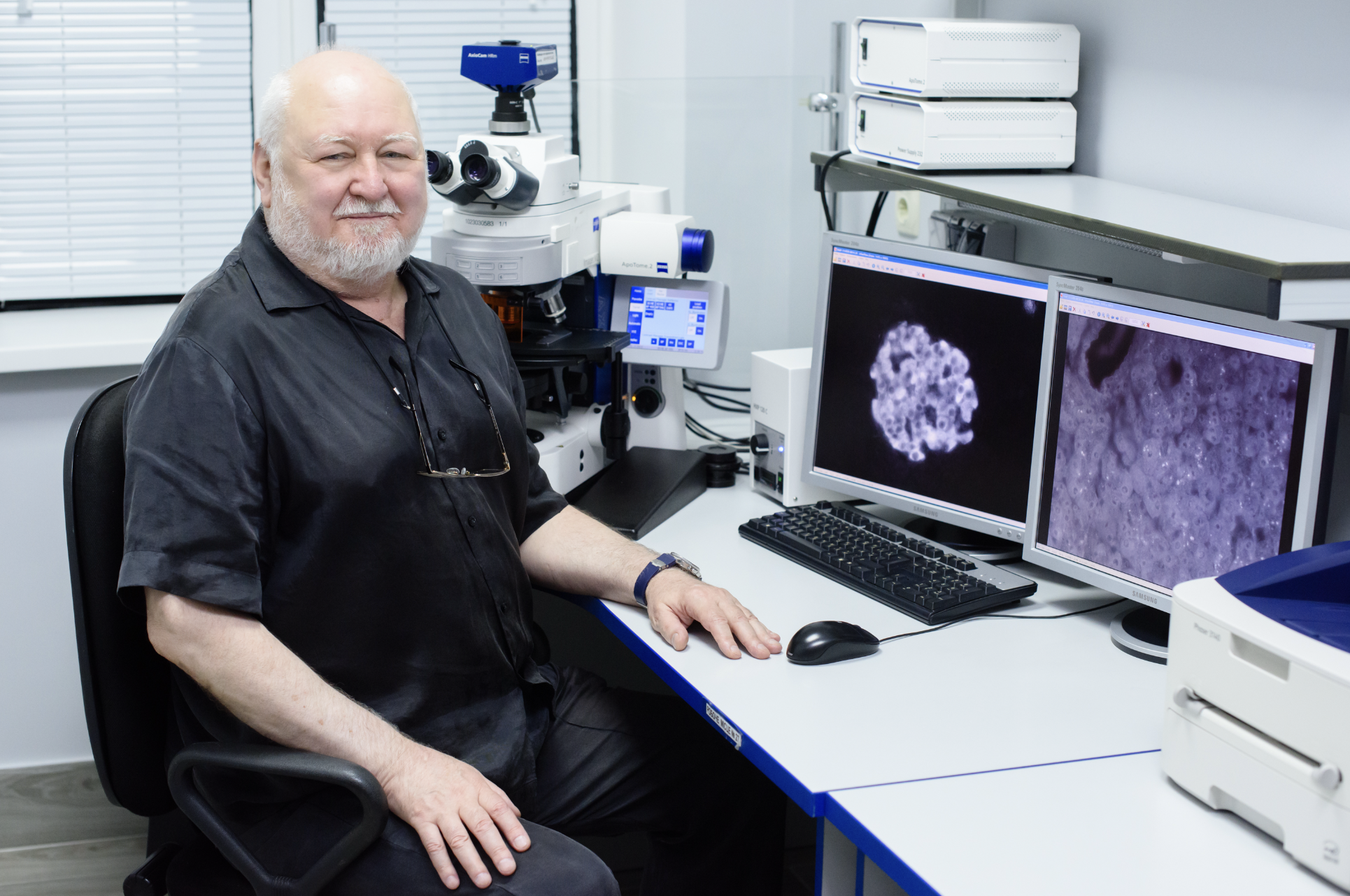
Prof. Giovanni Lombardi, from IRCCS Ospedale Galeazzi-Sant’Ambrogio in Milan (Italy) and Poznań University of Physical Education (Poland), has a strong scientific interest in the bone and muscle crosstalk, turnover and senescence, and is currently investigating the effects on bone microarchitecture of the exposure to perfluorononanoic acid (PFNA), a widely distributed yet under-studied PFAS, in rats, in collaboration with two groups of the CNR Institute of Agricultural Biology and Biotechnology (CNR-IBBA), based in Lodi and Pisa.
High-resolution micro-computed tomography (CT) analysis is a critical part of this study, as it provides data on bone microarchitecture necessary for downstream molecular analysis and for the subsequent identification of signalling pathways affected by PFNA.
For this reason, Prof. Lombardi requested access to the CNR Institute of Clinical Physiology in Pisa, part of our Multimodal Molecular Imaging Italian Node, taking advantage of the SEELIFE funding for user access which was available in 2025.
In Pisa, Wistar rats have been exposed to various doses of PFNA in drinking water over a 35-day period. At the end of the exposure period, histological evaluation and molecular and imaging analysis were conducted on the femurs and tibiae of the rats. This dual approach enabled a comprehensive assessment of both structural and molecular changes induced in bone tissue by PFNA exposure.
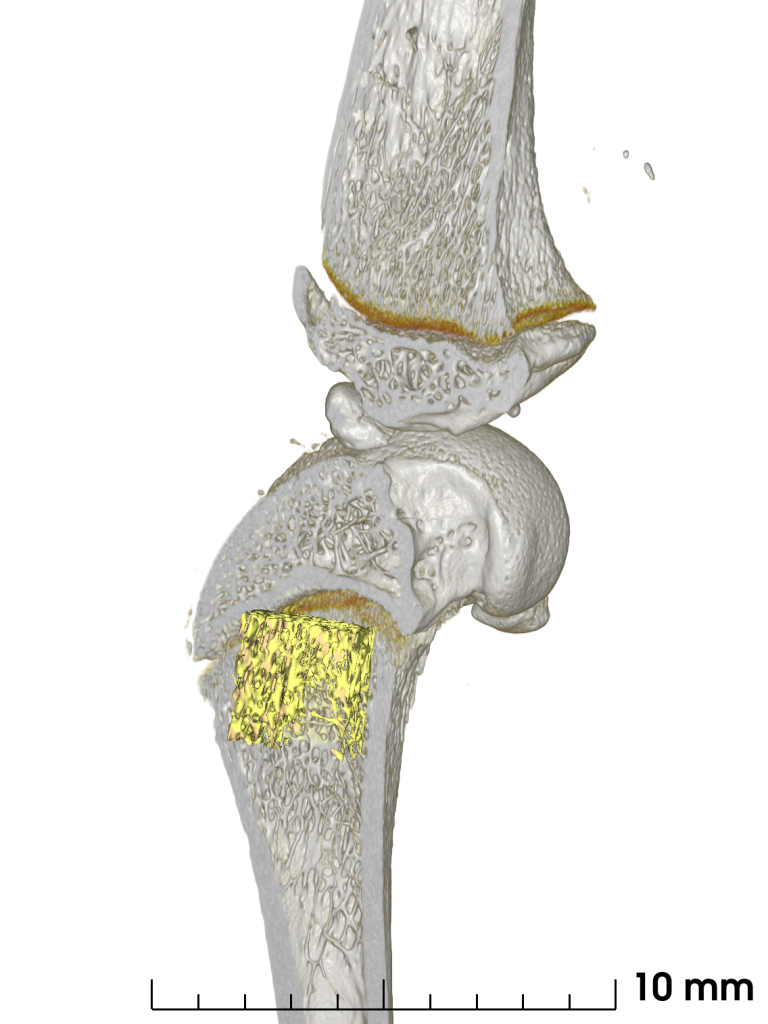
High-resolution CT was used to investigate the alterations in trabecular and cortical architecture of the femurs or tibiae, measuring trabecular thickness, trabecular spacing, bone volume fraction, connectivity density, cortical area, and bone mineral density, providing detailed insights into PFNA-induced alterations in bone microstructure.
Preliminary histological results have demonstrated that PFNA profoundly impacts bone structure and cellular organization. In particular, PFNA treatment results in a critical disorganization of the diaphyseal growth plate that may impact the osteoblast differentiation and, consequently, the structural organization of the bone. Furthermore, PFNA induces a relevant degree of fat substitution of the bone marrow and fat accumulation among the trabeculae. The analysis of the CT images is still ongoing: the extraction of parameters for both trabecular and cortical bone will clarify the negative effect of PFNA chronic exposure on bone microarchitecture. Specifically, Professor Lombardi and his team hypothesise that PFNA exposure results in decreased trabecular thickness and increased trabecular spacing, indicating a shift towards bone resorption and an aging-like phenotype.
The team suspects that these structural changes are dose-dependent, with the most severe disruption observed at the highest level of PFNA exposure. To confirm the hypothesis, additional measurements will be performed.
Conclusion and next steps
Employing a highly integrative and advanced methodological framework, including imaging technologies (CT), histopathological assessments, and molecular analyses, allowed to unravel structural, cellular, and molecular alterations in bones resulting from chronic PFNA exposure. Through this approach, the study contributed to elucidate novel mechanisms of PFNA action in the musculoskeletal system, thereby contributing to a broader understanding of PFAS toxicity and its relevance to public health.
The securing of SEELIFE funding, and the resulting opportunity to access the Euro-BioImaging imaging facilities – in particular the micro-CT – and to benefit from the expertise of a dedicated specialist team, offered us a unique opportunity to vastly expand the range of analyses we can perform on the available samples and, consequently, to extract as much information as possible. It therefore gave us the chance to explore aspects in greater depth that we would otherwise have been unable to address due to a lack of specific expertise within our research group, or for which it would have been necessary to incur costs that are not always sustainable.
– Professor Giovanni Lombardi
July 9, 2026
In April 2026, the Med-Hub Head of Operations Alessandra Viale attended the 2026 180°N Conference, held at the beautiful Nye Hjorten Teater…
July 7, 2026
Armed conflicts generate long-lasting environmental contamination that extends well beyond the duration of military operations. The release of heavy metals such as Arsenic,…

July 6, 2026
On 2 July 2026, Euro-BioImaging hosted the online EVOLVE workshop “Building High-Quality Preclinical Imaging Facilities”, bringing together approximately 40 imaging facility staff and…