The major impact of the coronavirus COVID-19 - including lockdown and quarantine in most parts of Europe - has changed the way scientists work. Laboratories around the world have shut down, and are required to develop new methods/processes to go back to work while respecting stringent hygiene measures. Members of Euro-BioImaging, a consortium of European countries and EMBL that provides life scientists with open access to a broad range of biological and biomedical imaging technologies, are implementing virtual and remote access to imaging infrastructure. Gleb Grebnev, the Manager of the Finnish Advanced Light Microscopy Node, a Euro-BioImaging Node in Finland, based in Turku, shares some preliminary exciting feedback from a successful virtual microscopy experience using STED microscope.
Tell us about the experiment you conducted with virtual microscopy and remote access? Gleb Grebnev: We looked at the fixed samples provided to us beforehand by a couple of users who normally come to our facility and image the samples themselves, but in the current situation were unable to do so. We looked at Mouse Embryonic Fibroblast cells under the super-resolution Abberior STED microscope. The samples were dual-labeled for vimentin and vinculin (focal adhesions) with Abberior STAR dyes. We used 100x objective for this purpose.
What technologies do you use for providing virtual microscopy to your imaging infrastructure? Gleb Grebnev: We have tested virtual microscopy on our STED machine, but in principle, the same set-up can be applied to all our microscopes. For screen sharing and voice, we use Zoom because both of our universities in Turku have licenses. Zoom also provides some functions that help the scientist guide the microscope operator with regards to selecting a region of interest by annotating the region on the screen.
What did you learn from the first experience? Gleb Grebnev: Sample selection prior to virtual microscopy is the key to success. Samples can be checked beforehand by an operator for levels of fluorescence. Remote users can also provide reference images, either their own or from publications so that an operator has a clear idea of what to look out for. We found that it works best for the scientist to send several samples by mail to the lab ahead of time so the microscope operator can choose the best one based on the scientist’s needs. The next difficulty is the selection of a focal plane, especially when it comes to thick samples. This depends not only on the competence of a microscope operator but also and on the resolution of a remote user’s screen.
What are some limitations to be aware of? Gleb Grebnev: On the remote user’s end, there is a slight delay, for example, when the sample is being focused. This is natural, but it requires remote users to be patient. Likewise, there is a delay in certain operations when a user accesses image acquisition software remotely. In addition, there is a considerable amount of background noise (e.g. microscope components, fans, ventilation), so a good microphone and good head/earphones are important. The virtual imaging session is longer than if a user accesses a microscope directly. Finally, the selection of the field of view with eyes is only available to the microscope operator and not a remote user. This is very critical as finding the most representative region in a sample is of crucial importance. Reference images come in handy here as well.
What feedback – if any- do you have from your user? Gleb Grebnev: The session went smoothly, the user was able to obtain good STED images from 5 different fields of view, and the whole process took about an hour. Check out the video to get a feel for what virtual microscopy is like.
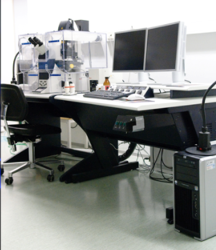
Above: Euro-BioImaging’s Finnish Advanced Light Microscopy Node shares its experience providing virtual access to its Abberior STED microscope with users Mayank Modi and Arun Venu from Åbo Akademi University and Turku Bioscience.
How do you make the images available to the user? Gleb Grebnev: Currently, for all of our remote users including international users, we upload the image data to our Seafile cloud storage provided by the University of Turku IT services and share the link with a remote user after which remote users download their data. OMERO server for data management and sharing is currently in the works to improve our data management capabilities.
Perspectives The Finnish Advanced Light Microscopy Node is currently inviting proposals for remote access projects from Finnish and international Euro-BioImaging users and is pleased to provide virtual access to its microscopes including the STED microscope mentioned in this story. This is a great way to move the research forward.
More news from Euro-BioImaging
July 15, 2026
Imaging-Driven Neuroscience, Responsible Research, and the Power of Advocacy: Highlights from FENS Forum 2026
Euro-BioImaging was pleased to participate in the FENS Forum 2026, Europe’s largest international gathering dedicated to advancing neuroscience (>8000 participants). Throughout…
Euro-BioImaging Highlights the Value of Research Infrastructures at the FENS 2026 Satellite Symposium hosted by EBRAINS
Euro-BioImaging was pleased to participate #FENS2026 Satellite Symposium “Accelerating Your Neuroscience Research through the European Research Infrastructures” organised by EBRAINS, to learn…